Thursday, 2 November 2017

Microbiology Week 7 and Experiments

      Assalmualaikum and good afternoon everyone. Hw r u? I hope you all fine as well. 😁😁😁 This week is my seventh week of microbiology class and this week I like to combine my post of microbiology and my basic technique of microbiology because they are interconnected with each other. In my basic technique class we did experiments 9 which is Gram staining and experiment 10 which is Acid fast staining. In my microbiology class, on wednesday we all having our suspend quiz on that but it is not individual quiz, it is team quiz about intracellular structure of prokaryotes  πŸ˜”πŸ˜”πŸ˜”and today we are going to study about intracellular structure of prokaryotes.


Basic techniques of microbiology 

EXPERIMENT 9 : Gram staining and EXPERIMENT 10: Acid fast staining
   
    After we enter the class, Dr Adelene talked about our test and what the mistake we did in our test. Then, Dr Adelene give some briefing and introduction about the experiment 9 and 10. For a change, this week Encik Zainuddin didn't give some demo how to do this two experiments. We did the experiments by our ownself. And after we finished our experiments, my demo was talked about the mistakes of the previous lab report. Half of my classmate did wrong in experiment A which is about measurement of microorganisms and again she taught us how to calculate it. So now I'm going to share the information about what I got learned in this week about experiment 9 and 10. 
                                                                   Before staining
After staining 


Experiemnt 9 : Gram staining

 The Gram stain is the most important stain procedure in microbiology. It is used to differentiate between gram positive organisms and gram negative organisms. Hence, it is a differential staining.  Differential staining requires the use of at least four chemical reagents that are applied sequentially to a heat-fixed smear. The first reagent is called primary stain is to impart it's color to all cells. The second stain is a mordant used to intensify the color of the primary stain. Based on the chemical composition of cellular components, the decolorizing agent may or may not remove the primary stain from the entire cell or only from certain cell structures. The final reagent is the counterstain, has a contrasting color to that of the primary stain. Gram stain reaction is based on the difference in the chemical composition of bacterial cell walls. Gram positive cell have thick layer of peptidoglycan but gram negative have thin layer of peptidoglycan and surrounded by outer lipid-containing layers. Peptidoglycan is mainly a polysaccharides composed of two chemical subunits found only in the bacterial cell wall. These subunits are N-acetylglucosamine and N-acetylmuramic acid.

Primary stain
    The primary stain is used in Gram staining to detect the Gram positive bacteria. The primary stain is a crystal violet color. When a bacteria is Gram negative, it loses the primary stain and takes on the counterstain of safranin. The violet stain is used first and stains all cells purple.

Mordant
    A mordant is a chemical used to hold down molecules of a stain onto a microorganism. Classically defined, mordants are usually ions such as metal ions or halide ions, but can be any molecule that serves the purpose of holding down a dye. However, a molecule called phenol is a non-ionic mordant that is discussed below. Some mordants bind both the dye and proteins on the microorganism. Most mordants are ions because the electrical charge on the ion attracts the electrical charge on a chemical dye. Thus, when the ion binds the dye, they form a large complex that precipitates -- meaning they become a solid and are no longer dissolved in the solution. Mordants hold down, or weigh down, the dye so it does not wash away during the remainder of the staining procedure. Washing is done so that only the true staining regions are visualized. At this point, all cells appear purple-black.

Decolorizing agent
    Alcohol or acetone dissolves the lipid outer membrane of Gram negative bacteria, thus leaving the peptidoglycan layer exposed and increases the porosity of the cell wall. The CV-I complex is then washed away from the thin peptidoglycan layer, leaving Gram negative bacteria colorless. On the other hand, alcohol has a dehydrating effect on the cell walls of Gram positive bacteria which causes the pores of the cell wall   to shrink. The CV-I complex gets tightly bound into the multi-layered, highly cross-linked Gram positive cell wall thus staining the cells purple. The decolorization step must be performed carefully, otherwise over-decolorization may occur. This step is critical and must be timed correctly otherwise the crystal violet stain will be removed from the Gram-positive cells. If the decolorizing agent is applied on the cell for  too long time , the Gram-positive organisms to appear Gram-negative. Under-decolorization occurs when the alcohol is not left on long enough to wash out the CV-I complex from the Gram-negative cells, resulting in Gram-negative bacteria to appear Gram-positive. Thus, the tightly bound primary stain complex is difficult to remove, and the cells remain purple.

Counterstain
    The decolorized Gram negative cells can  be rendered visible with a suitable counterstain, which is usually positively charged safranin, which stains them pink. Pink colour which adheres to the Gram positive bacteria is masked by the purple of the crystal violet.

Experiment 10: Acid fast staining

     The acid-fast stain is a differential stain used to identify acid-fast organisms such as members of the genus Mycobacterium. Acid-fast organisms are characterized by wax-like, nearly impermeable cell walls; they contain mycolic acid and large amounts of fatty acids, waxes, and complex lipids.  Acid-fast organisms are highly resistant to disinfectants and dry conditions. Because the cell wall is so resistant to most compounds, acid-fast organisms require a special staining technique. The primary stain used in acid-fast staining, carbolfuchsin, is lipid-soluble and contains phenol, which helps the stain penetrate the cell wall. This is further assisted by the addition of heat. The smear is then rinsed with a very strong decolorizer, which strips the stain from all non-acid-fast cells but does not permeate the cell wall of acid-fast organisms. The decolorized non-acid-fast cells then take up the counterstain. In our lab, we use the Ziehl-Neelsen method but not the Kinyoun method.

Primary stain
    Carbol fuchsin is a dark red stain in 5% phenol that is soluble in the lipoidal materials that constitute most of the mycobacterial cell wall, does penetrate these bacteria and its retained. Penetration is further enhanced by the application of heat, which drives the carbol fuchsin through the lipoidal wall and into the cytoplasm. This appllication of heat is used in the Ziehl-Neelsen method. The Kinyoun method, a modification of the Ziehl-Neelsen method, a modification of the Ziehl-Neelsen method, circumvents the use of heat by addition of a wetting agent to this stain, which reduces surface tension between the cell wall of the mycobacteria and the stain. Following application of the primary stain, all cells will appear red.

Decolorizing agent
      On application of acid alcohol (3% HCL + 95% Ethanol), the acid-fast cell will be resistant to decolorization, since the primary stain is more soluble in the cellular waxes than the decolouring reagent. In this event, the primary stain is retained and the mycobacteria will stay red. This is not the case with the nonacid-fast organisms that lacks the cellular waxes in their cell wall. The primary stain is more easily removed during decolorization, leaving these cells colourless or unstained.

Counterstain
Methylene blue is used as the final reagent to stain previously depolarized cell. As only nonacid-fast cells undergo decolourization, they may now absorb the counter stain and take on its blue color, while acid-fast cells retain the red color of the primary stain.
                                                 
                                  Staphylococcus aureus in Gram stain
                                                               

  Mixture of Mycobacterium smegmatis and Staphylococcus aureus in Acid fast stain

                                 Mycobacterium smegmatis in Acid fast stain

Mixture of Escherichia coli and Bacillus cereus in Gram stain


Microbiology Week 7 ( Intracellular structures of prokaryotes)
   
      The intracellular structures of prokaryotes which are plasma membrane, cytoplasm, the nuclear area, ribosomes, inclusions and endospore. Plasma membrane structure is phospholipid bilayer with proteins embedded in and attached to inner and outer surfaces. The function of the plasma membrane is selectively permeable barrier which is phospholipids are liquid at body temperature functions as a Fluid Mosaic, synthesizes cell wall components, assists in DNA replication, carries on respiration and captures energy as ATP. Destruction of the plasma membrane is disinfectants which is alcohol and quaternary ammonium compunds cause leakage of intracellular contents. The movement of materials across membranes is passive processes and active processes. Passive processes is molecules cross the membrane from an area of high concentration to an area of low concentration and the concentration gradient not energy dependent. Passive processes can classify into three processes which is simple diffusion, facilitated diffusion and osmosis. Simple diffusion is movement of molecules or ions from high to low concentration until equilibrium. Facilitated diffusion is a substances are moved by transporter proteins from high to low concentration and  needs a carrier proteins as transporters. Osmosis is movement of water molecule from high to low concentration. Active process is energy dependent system and ATP or proton motive force used. Active process can classify into two process which is active transport and group translocation. Active transport is substances are moved by transporter proteins from low to high concentration and cell has to expend energy for this to happen. Group translocation is molecules are chemically modified during passage across cytoplasmic membrane and energy is expended.
       Cytoplasm contains proteins (enzymes), carbohydrates, lipids, inorganic ions and many low molecular weight compounds. The major structures in the prokaryotic cytoplasm are DNA, ribosomes and inclusions. In intracytoplasmic membranes, plasma membrane infoldings is observed in many photosynthetic bacteria and observed in many bacteria with high respiratory activity and Anammoxosome in Planctomycetes is the organelle is site of anaerobic ammonia oxidation.
       The nuclear area is single long circular molecule of double-stranded DNA (bacterial chromosome). Bacterial chromosome do not include histones and are not sirrounded by nuclear envelope. In addition to the bacterial chromosome, bacteria often contain small circular, double-stranded DNA molecules called plasmid. Plasmid is a small circular, and double-stranded DNA. Extrachromosomal genetic elements not connected to bacterial chromosome and replicate independently of chromosomal DNA. Plasmid do not contain genetic material essential for growth. Plasmid is also contains features that enhance survivability examples of gene for drug resistance and it is also transferable from one bacterial to another.
      The function of the ribosome is sites of protein synthesis. It is consists of 70S ribosome. Ribosome has two subunit (small subunit-30S subunit and large subunit- 50S subunit). S refer to Svedberg unit. Each subunit consists of protein and RNA called ribosomal RNA or rRNA. Ribosome can be inhibited by certain antibiotics. Ribosome's cells that have high rates of protein synthesis, have a large number of ribosomes. Several antibiotics work by inhibiting protein synthesis on prokaryotic ribosomes which is streptomycin and gentamicin attach to the 30S subunit and erythromycin and chloramphenical attach to the 50S subunit.
    Inclusions is reserve deposits and can serve as a basis of identification. The type of inclusions are metachromatic granules, polysaccharide granules, lipid inclusions, sulfur granules, carboxysomes, magnetosomes and gas vacoule. Metachromatic granules is the large inclusion and stain red with certain blue dyes such as methylene blue. Metachromatic granules collectively known volutin is the inorganic phosphate and the phosphate used in the synthesis of ATP. Polysaccharide granules consist of glycogen and starch. Differentiated of polysaccharide granules by using iodine, reddish brown indicates glycogen granules and blue indicates starch granules. Lipid granules is storage material which is polymer poly-Ξ²-hydroxybutyric acid and revealed by Sudan dyes (fat-soluble). Sulfur granules is serves as an energy reserve. Derive energy by oxidising sulfur and sulfur containing compunds. Carboxysomes is contain the enzyme ribulose 1,5-diphosphate carboxylase and used for carbon dioxide fixation during phtosynthesis. Magnetosomes is a iron oxide that act like magnets and for downward movement until reaching suitable attachment site. Function of magnetosomes is to protect the cell against hydrogen peroxide accumulation. Gas vesicles is a hallow cylinder covered by proteins and collectively called gas vacuole. Gas vesicle appeared bright, refractile areas with an irregular outline in the phase microscope and consists of thin membrane surrounding a hallow space. Function of gas vesicles is to provide buoyancy for aquatic prokaryotes to receive sufficient amounts of oxygen, light and nutrients.
      Endospore is a resting structures formed by some bacteria for survival during adverse environmental conditions and germination results in leaving the dormant stage and once again becoming a typical, multiplying cell (vegetative cell). Genus Bacillus and Clostridium are two common disease causing bacteria that produce endospores as needed. Endospores cannot be destroyed easily, even by harsh chemicals, are formed internal to the bacterial cell membrane, highly resistant differentiated bacterial cell, and enable the organism to endure extreme environmental conditions. Endospore is a formation leads to a highly dehydrated structure thick walls and additional layers and contain essential macromolecules and a variety of substances absent from vegetative cells. Endospore called because the spore is formed within the cell. Endospores's spores are very impermeable to dye. However, they can be stained with special dye, Malachite green. Endospore is also small acid-soluble proteins to protect DNA from UV radiation, desiccation, and dry heat and serve as a carbon and energy source during germination. Endospore can remain dormant indefinitely but germinate quickly when the appropriate trigger is applied. Endospores and Infectious disease, although harmless themselves until they germinate, they are involved in the transmission of some disease to humans. Infections transmitted to humans by endospores included Bacillus anthracis, Clostridium tetani, Clostridium botulinum, and Clostridium perfringens.  
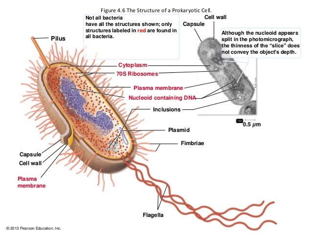
   Image result for intracellular structures of prokaryotes


Monday, 30 October 2017

Microbiology Week 6

    Assalamualaikum and good evening to everyone that read my post.... Hw r u everyone? I hope everyone will fine. 😁😁😁 This week is my sixth week of microbiology class and my test 1 is also in this week. πŸ˜‘πŸ˜‘πŸ˜‘ Everyweek have two microbiology but this week only one microbiology class because in another class, we all did out test 1 of microbiology. The test quiet difficult not all questions are difficult. There are some of the questions are easy. Usually on wednesday is my  microbiology class, but for change my microbiology class on tuesday. On tuesday, we learned about introduction to prokaryotes. The class start at 12 to 1pm. That time usually I'm so sleepy 😴😴😴 and I undrrstand a bit about the topic. Dr Wan also quiet fast when taught us that day. So now, I'm going to share about what I learned for this week.

    The prokaryotes are a group of organisms that lack a cell nucleus or any other membrane-bound organelles. Most are unicellular, but a few prokaryotes such as myxobacteria have multicellular stages in their life cycles. The size of prokaryotes is 0.2 to 2.0ΞΌm in diameter and 2 to 8ΞΌm in length. There are three basic shapes which are coccus, bacillus and spiral. Examples of cocci are diplococci, staphylococci, and streptococci. Examples of bacilli are diplobacilli and streptobacilli. Examples of spiral are vibrio, spirillum and spirochetes. Diplococci is in pairs, streptococci is in chain, staphylococci is grape-like cluster, tetrads is four cocci in a square and sarcinae in cubic configuration of eight cocci. The structures external to the cell wall is glycocalyx, flagella, axial filaments and fimbriae and pili. Glycocalyx is substance that surround cell and it is made inside the cell and excreted to the cell surface. The function of glycocalyx is protection from phygocytosis, attachment to various surfaces, source of nutrients and protect a cell against dehydration. Flagella is threadlike, locomotor appendages extending outward from plasma membrane and cell wall. Th e function of flagella is motility and swarming behavior, attachment to surfaces and may be virulence factors. The axial filaments is bundle of fibrils that arise at the ends of the cell beneath the outer sheath. Fimbriae and pili is hairlike appendages that are shorter, straighter and thinner than flagella. It consist of a protein called pilin arranged helically around a central core.

    The cell wall surrounds the cytoplasmic membrane and not a regulatory structure like cytoplasmic membrane. Composition and characteristics of cell wall is composed of a macromolecular network called peptidoglycan. Peptidoglycan consists of a repeating disaccharide. The disaccharide portion is made up of monosaccharieds called N-acetylglucosamine (NAG) and N-acetylmuramic acid (NAM). Alternating NAM and NAG molecules are linked in row by glycosidic bonds and adjacent rows are linked by peptide bonds. Functions of cell wall is prevent bacterial cell from rupturing when the water pressure inside the cell is greater than that outside the cell, contributes to pathogenicity, maintain characteristics shape, provides a rigid platform and counters the effect of osmotic pressure. Cell wall have two major types of walls which are Gram-positive and Gram-negative. Gram positive cell walls consists of many layers of peptidoglycan. Periplasmic space of Gram positive bacteria lies between plasma membrane and cell wall and is smaller than that of Gram- negative bacteria. Teichoic acid is primarily of an alcohol and phosphate, negatively charged, provide much of the wall' s antigenic specificity and cell walls do not degrade as easily. Two classes of teichoic acids which are lipoteichoic acid and wall teichoic acid. Gram negative cell wall consists of one or very few layers of peptidoglycan . The peptidoglycan is bonded to lipoprotein and does not contain teichoic acid. Periplasmic contains a high concentration of degrading enzymes and large number of transport proteins. Gram-negative cell wall consist of outer membrane is outer membrane consists of lipoproteins, lipopolysaccharides, phospholipids and porins, strong negative charge, and provide a barrier to certain antibiotic, lysozyme, detergent, heavy metals, bile salts and certain dyes.

     Atypical cell walls is mycoplasma, chlamydiaceae and archaea. Mycoplasma cells have no walls or have very little wall material. It's membrane contain sterols, which impart rigidity to the membrane and can pass through most bacterial filters. Plasma membranes are unique in having lipids called sterols which protect them from osmotic lyses. Chlamydiaceae have two membrane, some genes for peptidoglycan synthesis found in genome and obligate intracellular parasites. Archaea are lack of peptidoglycan in their cell walls. Some species have cell wall consisting of polysaccharide, glycoprotein or protein but not peptidoglycan. However, contain a substance similar to peptidoglycan called pseupeptidoglycan. Most common wall type is paracrystalline surface layer (S-layer) made of protein or glycoprotein with hexagonal symmetry. S-layer surface, or outermost layer forming "lipidless membrane", found on Archaea a few in Gram-positive and Gram-negative, composed  of repeating subunits of protein or glycoprotein, protect against osmotic stress, pH and enzyme and aid in attachment and inhibit phagocytosis. Archaea are naturally resistant to lusozyme and penicillin.


Image result for prokaryote               Image result for prokaryote


Image result for mycoplasma       

Friday, 13 October 2017

Microbiology Week 5

        Assalamualaikum and good morning everyone... How are you all ? πŸ˜ƒπŸ˜ƒπŸ˜ƒ This week is my fifth week of microbiology class huhu.... But actually I not yet adapt the environment in University Putra Malaysia as a degree student of microbiology. This week many things happened in my microbiology class. Firstly, For two days continuously Dr Wan gave some " tazkirah" which means advice to us because in class we are like not more curious to know about the microbes and all the things related to microbes. And also we even didn't look at what my friends posted in Open learning MOOC about the interesting things which are about Helicobacter pylori and Microbial interactions in termite guts. So, Dr Wan asked to make a group and each group have to choose one bacteria and we have to present it in three minutes. For my group, we were chose Magnetotatic bacteria. The Magnetotatic bacteria are quiet interesting because it is about magnetic. Others group also were chose about Mycobacterium tuberculosis, Serratia marcescens, Propiniobacterium acnes, Alcanivorax borkumensis and Pseudomonas aeruginosa. We were actually presented about our bacteria on friday. Each group must present 3 minutes, but some group presented more than 3 minutes because they want us to be more clear about what they are presented. Everyone were presented in our microbiology class for first time.   In my group , we were separated our work like one persons find about the history of that bacteria another about the characteristics of bacteria that my group chose. It is like team work and it make more closer to each other. So now I'm going to share about what I had studied for this week.

 Methods of classifying  and identifying microorganisms

        Morphological characteristics is easy to study and analaysed. Characteristics studied include cell type , shape and size, cellular grouping , external structures and internal structures. Differential staining which are Gram stain is classifies into gram positive or negative, Acid-fast stain is only stain bacteria with waxy material in their cell walls, Negative staining for capsule is use India ink to provide a contrasting background, then stain with simple stain and capsules to not accept simple stain, thus appear as halos surrounding bacterial cell, Endospore staining is appear green within pink cells and Flagella staining are too thin and use a mordant and stain carbolfuchsin to coat the flagella until they are thick enough to be seen. Biochemical test to verify its metabolic activity examples are Phenol Red broth, Gelatin test, Lipase test, Starch hydrolysis, Motility test and Catalase test. Serology is the science that studies serum and immune responses that are evident in serum and the background are Bacteria (antigen) enter a host, antibody will be produced by the host. The antibody will combine with the antigen ( bacteria) and this precipitates the antigen. Two types of serology which are Enzyme-linked immunosorbent assay (ELISA) is fast and utilised a computer scanner to read result and performed in microtiter plate and Western blotting is protein from an unknown bacterium or virus are separated by electrophoresis and transferred to a nitrocellulose filter by blotting.  Phage typing is to determine which phage a bacterium is susceptible to bacteriophage cause lysis of bacteria that they infect and infect only particular species or even strains. Fatty acid profiles is bacteria synthesise a wide variety of fatty acids these fatty acids are constant for a particular species. DNA Base Composition is the base composition of a single species is a fixed property that can be used to reveal the degree of species relatedness. DNA Fingerprinting is use of restriction enzyme to produce banding pattern. Nucleic Acid Hybridization can be used to determine extent of similarity based on degree of reunion. Techniques that apply the principle of nucleic acid hybridization are sounthern blotting, DNA chips, Ribotyping and Ribosomal RNA Sequencing and Fluorescent In Situ Hybridization. Methods use to classify and identify microorganisms after various analyses are Dichotomous Keys used for identification based on successive questions and Cladograms is show evolutionary relationships among organisms and constructed based on rRNA sequences with the aid of software.

Mycobacterium tuberculosis

Mycobacterium tuberculosis discovered by Robert Koch in 1882. Mycobacterium tuberculosis is a non motile, acid-fast, obligate aerobe. The bacilli are 2-4 micrometer in length and have a very slow generation of time between 15 to 20 hours. The cell wall of the Mycobaterium is unique in that it is composed mainly of acidic waxes, specifically mycolic acids. Mycobacterium tuberculosis is usually resistant to drying and chemicals, contributing to the ease with which it is transmitted. Mycobacterium tuberculosis is ropelike structure with peptidoglycan and found abundantly in soil. The tuberculin skin test currently used to diagnose infection with Mycobacterium tuberculosis has poor diagnostic value, especially in geographic areas where the prevalence of tuberculosis is low or where the environment burden of saprophytic, nontuberculous mycobacteria is high.


Alcanivorax borkumensis

Alcanivorax borkumensis is an alkane-degrading marine bacterium which naturally propagates and becomes predominant in crude-oil-containing seawater when nitrogen and phosphorus nutrients are supplemented. They are currently thought to be the world's most important oil-degrading organisms. This bacteria is useful in treating oil spillage because it is an oil-eating bacteria. A borkumensis is a rod-shaped bacterium without flagella that obtains its energy primarily from consuming alkanes. It is aerobic meaning it uses oxygen to gain energy and it is halophilic, meaning it tends to live in environments that contain salt, such as salty ocean water. It is also Gram-negative which essentially means it has a relatively thin cell wall. It is also non-motile, however other organisms that appear to be in the same genus are motile through flagella.


Propionibacterium acnes

 Propionibacterium acnes is the relatively slow-growing, typically aerotolerant anaerobic, Gram-positive bacterium (rod) linked to the skin condition of acne. P. acnes bacteria live deep within follicles and pores, away from the surface of the skin. In these follicles, P. acnes bacteria use sebum, cellular debris and metabolic byproducts from the surrounding skin tissue as their primary sources of energy and nutrients. Elevated production of sebum by hyperactive sebaceous glands (sebaceous hyperplasia) or blockage of the follicle can cause P. acnes bacteria to grow and multiply. P. acnes bacteria secrete many proteins, including several digestive enzymes. These enzymes are involved in the digestion of sebum and the acquisition of other nutrients. They can also destabilize the layers of cells that form the walls of the follicle. The cellular damage, metabolic byproducts and bacterial debris produced by the rapid growth of P. acnes in follicles can trigger inflammation. This inflammation can lead to the symptoms associated with some common skin disorders, such as folliculitis and acne vulgaris. P. acnes bacteria are susceptible to a wide range of antimicrobial molecules, from both pharmaceutical and natural sources. Antibiotics are commonly used to treat infections caused by P. acnes. Acne vulgaris is the disease most commonly associated with P. acnes infection. The antibiotics most frequently used to treat acne vulgaris are erythromycin, clindamycin, doxycycline, and minocycline. Several other families of antibiotics are also active against P. acnes bacteria, including quinolones, cephalosporins, pleuromutilins, penicillins, and sulfonamides.


Thursday, 5 October 2017

Microbiology Week 4

   Assalamualaikum and good afternoon eveyone that reading my blog now.... πŸ˜ƒπŸ˜ƒπŸ˜ƒπŸ˜ƒ. This week is my forth week of microbiology class. I feel like it is my first class but actually my forth class with Dr Wan... You know time flying so fast. I would like to share you what I learned in the forth week of microbiology class with my spotting lecturer Dr Wan. Usually after my microbiology class, I feel so tired and sometimes I a bit stress because a lot of works and reseach have to do. That was as usual because I as a microbiology student I have to do more reasearch to know and find the types of microoorganisms. Like a scientist huhuhu 😍😍😍. I feel stress because Im not yet adapt the changes in university. I think it will takes a bit more time to adapt the changing because now only my enter to the degree. But for sure oneday i will change and become a good and profesional scientist .😎😎😎 So on wednesday, Dr Wan taught us about electron microscope and on friday we all had group discussion about the classification of organisms. Group discussion is like in one group we have to separate the topic among with our group members and understand about the topics. Then who are got the same topics to be discuss they are will combine and make a group. They discuss about what they are understand and do more research. Afterthat, only two people are in the group and the rest of all can go to another topic groups and the another group members will explain about what they are understand about their topics. The discussion a quiet fun and we all enjoyed but we does not have enough time to go to more groups but Dr Wan said we will study again in next week.This week for classifications of organisms are short details. After Dr Wan explain it then most probably next week I will share more about classifications of organisms. So, Im going to share the information about electron microscope.

              Electron Microscope

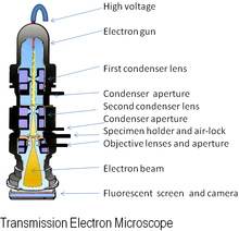
An electron microscope is a microscope that uses a beam of accelerated electrons as a source of illumination. As the wavelength of an electron can be up to 100, 000 times shorter than that of visible light photons, electron microscopes have a resolving power than light microscopes and can reveal the structure of smaller objects. Electron microscopes have electron optical lens systems that are analogous to the glass lenses of an optical light microscope.The similarity of scanning electron microscope (SEM) and transmission electron microscope (TEM) is are both structures smaller than 0.2 micrometer. The differences between of SEM and TEM are SEM produces a realistics 3D image of specimens's surface features but produces 2D image. The magnification of SEM is 1000X to 10,000X but in TEM are 10,000X to 100,000X. The resolving power for SEM is 20 nm but in TEM is 2.5 nm. SEM is to study the surface features of cells and viruses but TEM is to examine viruses or the internal ultrastructure in thin sections of cell. Electron Cryotomography is rapid technique provides way to preserve native state of structures examined in vacuum. Images recorded from many different directions to creates 3D structures.


Scanning Electron Microscope

view under electron microscope
         

Preparations of specimens for light microscopy 

Preparations of specimens for light microscopy are wet mount and smears. Smears-steps involve are preparing smears, fixation, and staining.Fixation is a preserves internal and external structures and fixed them in position. Stain is a salt composed of a positive and negative ion, one of which is colourful this will called the chromophore. Stain make internal and external structures of cell more visible by increasing contrast with background. There are three types of staining which are simple staining, differential staining and special staining. In differential staining have gram staining and acid-fast staining. In special staining have negative staining, endospore staining and flagella staining.

gram stain

acid-fast staining 

negative staining

endospore staining 

flagella staining 


Classification of organisms 

Taxonomy is a arrangement into groups based on mutual similarities. There are three interrelated parts in the taxonomy which classification, identification and nomenclature. Classification is the arrangement of organisms into groups or taxon based on mutual similarity or evolutionary relatedness. Identification is the process of discovering and recording the traits of organisms. Nomenclature is a assignment of names to taxonomic groups in agreement with published rules.Strain is a subgroup of species with one or more characteristics that distinguish it from other subgroups of the same species. Different ways to describe strains within a species which are biovars, morphovars and serovars. Phylogenetic classifications is a natural systems based on evolutionary relationships and direct comparison of genetic material and gene products.

Methods of classifying and identifying microorganisms

Methods of classifying and identifying microorganisms are morphological characteristics, differential staining, biochemical tests, serology, phage typing, fatty acid profiles, DNA base composition,DNA fingerprinting, nucleic acid hybridization. Methods use to classify and identify microorganisms after various analyses which are dichotomous keys and cladograms.


Saturday, 30 September 2017

Microbiology Week 3

Assalamualaikum and hello guys.... hw r u ?... I hope you all fine and great. I would like to share you what I learned in my third week of microbiology class with my energetic lecturer which is DR. Wan . My microbiology class is on Wednesday and Friday. For this week, on Wednesday Dr. Wan and my seniors went to the competition. And Dr. Wan got an award. Congratulations Dr. Wan. 😍😍😍So on Wednesday Dr. Wan gave a task to us in Open leaning MOOC. We all did the questions that related to microbiology. First of all I didn't understand the task as usual. But later on I can understand and did all the questions that Dr. Wan asked us to do. 😊😊😊 The task was like a bit game and it was actually interesting once you know how to play the task. In the task, there have a password and we have to find out the password. At last, we have to write the password in the space they provided. The task also help us to learn about microbiology and to revise what we had studied in past two weeks. I got the password of the task which is " Fungi Are Eukaryotes". πŸ˜…πŸ˜…πŸ˜… The open leaning MOOC is not only in my university which is University Putra Malaysia but they are a lot of universities and colleges around the world. It will help us to communicate with the people are in the foreign countries. We can also share about the microbes and about new microorganisms in the open learning MOOC page.

    On Friday, we have microbiology class with Dr. Wan in afternoon. Afternoon usually hot and I was really sleepy before I enter the class. 😴😴😴But once Dr. Wan enter our class, it will make me fresh because Dr. Wan make some fun and the way Dr teach us are quiet different from others. On Friday, I learned about the microscopy. πŸ”¬πŸ”¬πŸ”¬ The history of the microscopes

are the field of microscopy ( optical microscopy) has its roots in the 17th-century Dutch Republic, with some notable representatives such as Zacharias Janssen, Hans Lippershey, Jan Swammerdam, and Antonie van Leeuwenhoek. Van Leeuwenhoek is often considered to be the first acknowledged microscopist and microbiologist.

   Microscopy is the technical field of using microscopes to view objects and areas of objects that cannot be seen with the naked eye ( objects that are not within the resolution range of the normal eye). There are three well-known branches of microscopy which are optical, electron and scanning probe microscopy. Goals of microscopes are to produce a magnified image of the specimen, separate the details in the image, and render the details visible to the human eye or camera. Microscopy have two categories which are light microscopes and electron microscope. In light microscope have many varieties which are bright-field microscopes, dark-field microscopes, phase-contrast microscope, fluorescence microscopes and confocal microscope. I got learned about the parts of microscope which are ocular, body, nosepiece, objective lens, arm, mechanical stage, substage condenser, diaphragm, base with light source, field diaphragm lever, light intensity control, coarse focus adjustment knob, fine focus adjustment knob and the stage adjustment knobs. Bright-field microscope that use in lab requires stain and produce a dark image against a brighter background . Dark-field microscope image is formed by light reflected or refracted by specimen and produces a bright image of the object against a dark background. The phase-contrast microscope converts differences in refractive index into detected variations in light intensity. The phase-contrast is excellent way to observe living cells and stain is not necessary view internal structures of living organisms. The fluorescence microscopes exposes specimen to ultraviolet or blue light and the specimens usually stained with fluorochromes. Cell contains natural fluorescent substances example (chlorophyll) has been treated with fluorescent dye. The confocal microscopy is confocal scanning laser microscopy creates sharp, composite 3D image of specimens by using laser beam, aperture to eliminate stray light and computer interface. Confocal microscopy is the numerous applications including study of biofilms.


Friday, 22 September 2017

Microbiology week 2

 
CHARACTERISTS OF MIROORGANISMS

 Microbiology are the smallest organisms on Earth. In fact, the term microorganism literally means "microscopic organism." Microorganisms may be composed of prokaryotic or eukaryotic cells, and they may be single-celled or multicellular. Examples of microorganisms include algae, fungi, protozoa, bacteria and viruses. Prokaryotic microorganisms is prokaryotes arguably represent the earliest forms of life on Earth. They are broken up into two categories which are bacteria and archaea. A prokaryotic cell lacks a nucleus to hold the cell's DNA and lacks any sort of organized packaging or housing to hold the rest of the cell's machinery. Because prokaryotic cells lack this extra material, they are almost always smaller than other cell types; all prokaryotes are microorganisms, and they are almost single-celled. Eukaryotic cells are larger and more complex than prokaryotic cells. The DNA of a eukaryotic cell is neatly packaged within its nucleus, and there are several different structures that house the cellular machinery that can make eukaryotic microorganisms self-sufficient. The structures located in eukaryotic cells may include the endoplasmic reticulum, golgi apparatus, mitochondria, and ribosomes, as well as chloroplasts in photosynthetic cells. Examples of eukaryotic microorganisms include fungi, algae, protozoa and various microscopic parasitic worms.

     Bacteria are individual living cells. Bacteria cells are similar to your cells in many ways; yet, they also have distinct differences. Bacteria have many unique adaptations allowing them to live in many different environments. Bacteria are so small that they can only be seen with a microscope. When viewed under the microscope, they have three distinct shapes. Bacteria can be indentified by their shape which are bacillus are rod-shaped, coccus are sphere-shaped and spirillius are spiral-shaped. Bacteria are like eukaryotic cells in that they have cytoplasm, ribosomes, and a plasma membrane. Features that distinguish a bacterial cell from a eukaryotic cell include the circular DNA of nucleoid, the lack of membrane-bound organelles, the cell wall of peptidoglycan, and flagella. Besides that, Protozoa are eukaryotic microorganisms. Although they are often studiedin zoology courses, they are considered part of the microbial world because they are unicellular and microscopic. Characteristics of Protozoa are they do not have cell wall; some however,possess a flexible layer, a pellicle, or a rigid shell of inorganic material outside the cell membrane. They have the ability during their entire life cycle or part of it to move by locomotor organelles or by a gliding mechanism. They have heterotrophic mode of nutrition, whereby the free-living forms ingest particulates, such as bacteria, yeast, and algae, while the parasitic forms derive nutrients from the body fluids of their hosts.

    Moreover, Algae are eukaryotic organisms that have no roots, stems, or leaves but do have chlorophyll and other pigments for carrying out photosynthesis. Algae can be multicellular or unicellular. Cells are covered by a rigid cellulose cell wall. Algae are eukaryotic thallophytes and photoautotrophs. Algae reproduce either by vegetative, asexual or sexual method. Storage form of food in algae is starch. Algae are usually aquatic, either freshwater or marine and some are terrestrial. Furthermore, Fungi is any member of the group of eukaryotic organisms that includes microorganisms such as yeasts and molds, as well as the more familiar mushrooms. These organisms are classified as a kingdom, Fungi, which is separate from the other eukaryotic life kingdoms of plants and animals. Characteristics of fungi are fungi have cell walls are composed mainly of a carbohydrate called chitin, while plant cell walls are composed mainly of cellulose. Fungi are achlorophyllous, which means they lack of chlorophyll pigments present in the chloroplasts in plant cells and which are necessary for photosynthesis. The carbohydrate molecule used to store nergy in fungi is glycogen. Fungi are heterotrophs, which means that they obtain nutrients by absorption. Hyphae can grow and form a network called a mycelium. A spore is a unit of asexual reproduction, specifically a reproductive cell surrounded by a thick cell wall. Yeasts are unicellular fungi that do not produce hyphae. Fungi can reproduce both sexually and asexually.

    Lastly, Virus is a small infections agent that replicates only inside the living cells of other organisms. Viruses can infect all types of life forms, from animals and plants to mcroorganisms, including bacteria and archaea. Characteristics of viruses are enclosed in protective envelope. They have spikes, which helps them to attach to the host cell. Viruses are non cellular and do not respire, do not metabolize and do not grow but they do reproduce. They contain a protein coat called the capsid. They have a nucleic acid core containing DNA or RNA. Ribosome and enzymes are absent, which are needed for metabolism. Viruses are considered both as living and non living things, as viruses are inactive when they are present outside of host cells and are active in side of host cells.


Examples of Bacteria :


Examles of Protozoa:


Examples of Fungi :  


Examples of Algae:


Examples of Viruses :


HISTORY OF MICROBIOLOGY

   Historians are unsure who made the first observations of microorganisms, but the microscope was available during the mid-1600s, and an English scientist named Robert Hooke made key obsevations. He is reputed to have observed strands of fungi among the specimens of cells he viewed. In the 1670s and the decades thereafter, a Dutch merchant named Anton van Leeuwenhoek made careful observations of microscopic organisms, which he called animalcules. Until his death in 1723, van Leeuwenhoek revealed the microscopic world to scientists of the day and is regarded as one of the first to provide accurate descriptions of protozoa, fungi, and bacteria.

   After van Leeuwenhoek died, the study of microbiology did not develop rapidly because microscopes were rare and the interest in microorganisms was not high. In those years, scientists debated the theory of spontaneous generation, which stated that microorganisms arise from lifeless matter such as beef broth. This theory was disputed by Francesco Redi, who showed that fly maggots do not arise from decaying meat (as others believed) if the meat is covered to prevent the entry of flies. An English cleric named John Needham advanced spontaneous generation, but Lazzaro Spallanzani disputed the theory by showing that boiled broth would not give rise to microscopic forms of life.

  Louis Pasteur and the germ theory. Louis Pasteur worked in the middle and late 1800s. He performed numerous experiments to discover why wine and dairy product became sour, and he found that bacteria were to blame. Pasteur called attention to the important of microorganisms in everyday life and stirred scientists to think that if bacteria could make the wine "sick",the perhaps they could cause human illness.

  Pasteur had to disprove spontaneous generation to sustain his theory, and he therefore devised a series of swan-necked flasks filled with broth. He left the flasks of broth open to the air, but the flasks had a curve in the neck so that microorganisms would fail into the neck, not the broth. The flasks did not become contaminated (as he predicted they would not), and Pasteur's experiments put to rest the notion of spontaneous generation. His work also encouraged the belief that microorganisms were in the air and could cause disease. Pasteur postulated the germ theory of disease, which states that microorganisms are the causes of infectious disease. 
 
   Pasteur's attempts to prove the germ theory were unsuccessful. However, the German scientist Robert Koch provided the proof by cultivating anthrax bacteria apart from any other type of organism. He then injected pure cultures of the bacilli invariably caused anthrax. The procedures used by Koch came to be known as Koch's postulates. They provided a set of principles whereby other microorganisms could be related to other diseases.                                                                                              

Anton van Leeuwenhoek


Robert Hooke


John Needham


Lazzaro Spallanzani


Louis Pasteur


Robert Koch


Microbiology Semester 2 Week 14

              Assalamualaikum and hi everyone... how are you all? I hope everyone will be fine as well... On Tuesday, before our class start...