Basic techniques of microbiology
EXPERIMENT 9 : Gram staining and EXPERIMENT 10: Acid fast staining
After we enter the class, Dr Adelene talked about our test and what the mistake we did in our test. Then, Dr Adelene give some briefing and introduction about the experiment 9 and 10. For a change, this week Encik Zainuddin didn't give some demo how to do this two experiments. We did the experiments by our ownself. And after we finished our experiments, my demo was talked about the mistakes of the previous lab report. Half of my classmate did wrong in experiment A which is about measurement of microorganisms and again she taught us how to calculate it. So now I'm going to share the information about what I got learned in this week about experiment 9 and 10.
Before staining

After staining

Experiemnt 9 : Gram staining
The Gram stain is the most important stain procedure in microbiology. It is used to differentiate between gram positive organisms and gram negative organisms. Hence, it is a differential staining. Differential staining requires the use of at least four chemical reagents that are applied sequentially to a heat-fixed smear. The first reagent is called primary stain is to impart it's color to all cells. The second stain is a mordant used to intensify the color of the primary stain. Based on the chemical composition of cellular components, the decolorizing agent may or may not remove the primary stain from the entire cell or only from certain cell structures. The final reagent is the counterstain, has a contrasting color to that of the primary stain. Gram stain reaction is based on the difference in the chemical composition of bacterial cell walls. Gram positive cell have thick layer of peptidoglycan but gram negative have thin layer of peptidoglycan and surrounded by outer lipid-containing layers. Peptidoglycan is mainly a polysaccharides composed of two chemical subunits found only in the bacterial cell wall. These subunits are N-acetylglucosamine and N-acetylmuramic acid.
Primary stain
The primary stain is used in Gram staining to detect the Gram positive bacteria. The primary stain is a crystal violet color. When a bacteria is Gram negative, it loses the primary stain and takes on the counterstain of safranin. The violet stain is used first and stains all cells purple.
Mordant
A mordant is a chemical used to hold down molecules of a stain onto a microorganism. Classically defined, mordants are usually ions such as metal ions or halide ions, but can be any molecule that serves the purpose of holding down a dye. However, a molecule called phenol is a non-ionic mordant that is discussed below. Some mordants bind both the dye and proteins on the microorganism. Most mordants are ions because the electrical charge on the ion attracts the electrical charge on a chemical dye. Thus, when the ion binds the dye, they form a large complex that precipitates -- meaning they become a solid and are no longer dissolved in the solution. Mordants hold down, or weigh down, the dye so it does not wash away during the remainder of the staining procedure. Washing is done so that only the true staining regions are visualized. At this point, all cells appear purple-black.
Decolorizing agent
Alcohol or acetone dissolves the lipid outer membrane of Gram negative bacteria, thus leaving the peptidoglycan layer exposed and increases the porosity of the cell wall. The CV-I complex is then washed away from the thin peptidoglycan layer, leaving Gram negative bacteria colorless. On the other hand, alcohol has a dehydrating effect on the cell walls of Gram positive bacteria which causes the pores of the cell wall to shrink. The CV-I complex gets tightly bound into the multi-layered, highly cross-linked Gram positive cell wall thus staining the cells purple. The decolorization step must be performed carefully, otherwise over-decolorization may occur. This step is critical and must be timed correctly otherwise the crystal violet stain will be removed from the Gram-positive cells. If the decolorizing agent is applied on the cell for too long time , the Gram-positive organisms to appear Gram-negative. Under-decolorization occurs when the alcohol is not left on long enough to wash out the CV-I complex from the Gram-negative cells, resulting in Gram-negative bacteria to appear Gram-positive. Thus, the tightly bound primary stain complex is difficult to remove, and the cells remain purple.
Counterstain
The decolorized Gram negative cells can be rendered visible with a suitable counterstain, which is usually positively charged safranin, which stains them pink. Pink colour which adheres to the Gram positive bacteria is masked by the purple of the crystal violet.
Experiment 10: Acid fast staining
The acid-fast stain is a differential stain used to identify acid-fast organisms such as members of the genus Mycobacterium. Acid-fast organisms are characterized by wax-like, nearly impermeable cell walls; they contain mycolic acid and large amounts of fatty acids, waxes, and complex lipids. Acid-fast organisms are highly resistant to disinfectants and dry conditions. Because the cell wall is so resistant to most compounds, acid-fast organisms require a special staining technique. The primary stain used in acid-fast staining, carbolfuchsin, is lipid-soluble and contains phenol, which helps the stain penetrate the cell wall. This is further assisted by the addition of heat. The smear is then rinsed with a very strong decolorizer, which strips the stain from all non-acid-fast cells but does not permeate the cell wall of acid-fast organisms. The decolorized non-acid-fast cells then take up the counterstain. In our lab, we use the Ziehl-Neelsen method but not the Kinyoun method.
Primary stain
Carbol fuchsin is a dark red stain in 5% phenol that is soluble in the lipoidal materials that constitute most of the mycobacterial cell wall, does penetrate these bacteria and its retained. Penetration is further enhanced by the application of heat, which drives the carbol fuchsin through the lipoidal wall and into the cytoplasm. This appllication of heat is used in the Ziehl-Neelsen method. The Kinyoun method, a modification of the Ziehl-Neelsen method, a modification of the Ziehl-Neelsen method, circumvents the use of heat by addition of a wetting agent to this stain, which reduces surface tension between the cell wall of the mycobacteria and the stain. Following application of the primary stain, all cells will appear red.
Decolorizing agent
On application of acid alcohol (3% HCL + 95% Ethanol), the acid-fast cell will be resistant to decolorization, since the primary stain is more soluble in the cellular waxes than the decolouring reagent. In this event, the primary stain is retained and the mycobacteria will stay red. This is not the case with the nonacid-fast organisms that lacks the cellular waxes in their cell wall. The primary stain is more easily removed during decolorization, leaving these cells colourless or unstained.
Counterstain
Methylene blue is used as the final reagent to stain previously depolarized cell. As only nonacid-fast cells undergo decolourization, they may now absorb the counter stain and take on its blue color, while acid-fast cells retain the red color of the primary stain.
Staphylococcus aureus in Gram stain

Mixture of Mycobacterium smegmatis and Staphylococcus aureus in Acid fast stain

Mycobacterium smegmatis in Acid fast stain

Mixture of Escherichia coli and Bacillus cereus in Gram stain

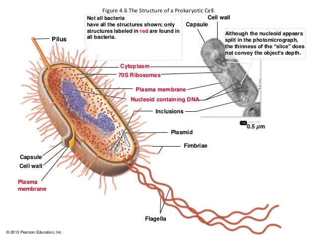
Microbiology Week 7 ( Intracellular structures of prokaryotes)
The intracellular structures of prokaryotes which are plasma membrane, cytoplasm, the nuclear area, ribosomes, inclusions and endospore. Plasma membrane structure is phospholipid bilayer with proteins embedded in and attached to inner and outer surfaces. The function of the plasma membrane is selectively permeable barrier which is phospholipids are liquid at body temperature functions as a Fluid Mosaic, synthesizes cell wall components, assists in DNA replication, carries on respiration and captures energy as ATP. Destruction of the plasma membrane is disinfectants which is alcohol and quaternary ammonium compunds cause leakage of intracellular contents. The movement of materials across membranes is passive processes and active processes. Passive processes is molecules cross the membrane from an area of high concentration to an area of low concentration and the concentration gradient not energy dependent. Passive processes can classify into three processes which is simple diffusion, facilitated diffusion and osmosis. Simple diffusion is movement of molecules or ions from high to low concentration until equilibrium. Facilitated diffusion is a substances are moved by transporter proteins from high to low concentration and needs a carrier proteins as transporters. Osmosis is movement of water molecule from high to low concentration. Active process is energy dependent system and ATP or proton motive force used. Active process can classify into two process which is active transport and group translocation. Active transport is substances are moved by transporter proteins from low to high concentration and cell has to expend energy for this to happen. Group translocation is molecules are chemically modified during passage across cytoplasmic membrane and energy is expended.
Cytoplasm contains proteins (enzymes), carbohydrates, lipids, inorganic ions and many low molecular weight compounds. The major structures in the prokaryotic cytoplasm are DNA, ribosomes and inclusions. In intracytoplasmic membranes, plasma membrane infoldings is observed in many photosynthetic bacteria and observed in many bacteria with high respiratory activity and Anammoxosome in Planctomycetes is the organelle is site of anaerobic ammonia oxidation.
The nuclear area is single long circular molecule of double-stranded DNA (bacterial chromosome). Bacterial chromosome do not include histones and are not sirrounded by nuclear envelope. In addition to the bacterial chromosome, bacteria often contain small circular, double-stranded DNA molecules called plasmid. Plasmid is a small circular, and double-stranded DNA. Extrachromosomal genetic elements not connected to bacterial chromosome and replicate independently of chromosomal DNA. Plasmid do not contain genetic material essential for growth. Plasmid is also contains features that enhance survivability examples of gene for drug resistance and it is also transferable from one bacterial to another.
The function of the ribosome is sites of protein synthesis. It is consists of 70S ribosome. Ribosome has two subunit (small subunit-30S subunit and large subunit- 50S subunit). S refer to Svedberg unit. Each subunit consists of protein and RNA called ribosomal RNA or rRNA. Ribosome can be inhibited by certain antibiotics. Ribosome's cells that have high rates of protein synthesis, have a large number of ribosomes. Several antibiotics work by inhibiting protein synthesis on prokaryotic ribosomes which is streptomycin and gentamicin attach to the 30S subunit and erythromycin and chloramphenical attach to the 50S subunit.
Inclusions is reserve deposits and can serve as a basis of identification. The type of inclusions are metachromatic granules, polysaccharide granules, lipid inclusions, sulfur granules, carboxysomes, magnetosomes and gas vacoule. Metachromatic granules is the large inclusion and stain red with certain blue dyes such as methylene blue. Metachromatic granules collectively known volutin is the inorganic phosphate and the phosphate used in the synthesis of ATP. Polysaccharide granules consist of glycogen and starch. Differentiated of polysaccharide granules by using iodine, reddish brown indicates glycogen granules and blue indicates starch granules. Lipid granules is storage material which is polymer poly-β-hydroxybutyric acid and revealed by Sudan dyes (fat-soluble). Sulfur granules is serves as an energy reserve. Derive energy by oxidising sulfur and sulfur containing compunds. Carboxysomes is contain the enzyme ribulose 1,5-diphosphate carboxylase and used for carbon dioxide fixation during phtosynthesis. Magnetosomes is a iron oxide that act like magnets and for downward movement until reaching suitable attachment site. Function of magnetosomes is to protect the cell against hydrogen peroxide accumulation. Gas vesicles is a hallow cylinder covered by proteins and collectively called gas vacuole. Gas vesicle appeared bright, refractile areas with an irregular outline in the phase microscope and consists of thin membrane surrounding a hallow space. Function of gas vesicles is to provide buoyancy for aquatic prokaryotes to receive sufficient amounts of oxygen, light and nutrients.
Endospore is a resting structures formed by some bacteria for survival during adverse environmental conditions and germination results in leaving the dormant stage and once again becoming a typical, multiplying cell (vegetative cell). Genus Bacillus and Clostridium are two common disease causing bacteria that produce endospores as needed. Endospores cannot be destroyed easily, even by harsh chemicals, are formed internal to the bacterial cell membrane, highly resistant differentiated bacterial cell, and enable the organism to endure extreme environmental conditions. Endospore is a formation leads to a highly dehydrated structure thick walls and additional layers and contain essential macromolecules and a variety of substances absent from vegetative cells. Endospore called because the spore is formed within the cell. Endospores's spores are very impermeable to dye. However, they can be stained with special dye, Malachite green. Endospore is also small acid-soluble proteins to protect DNA from UV radiation, desiccation, and dry heat and serve as a carbon and energy source during germination. Endospore can remain dormant indefinitely but germinate quickly when the appropriate trigger is applied. Endospores and Infectious disease, although harmless themselves until they germinate, they are involved in the transmission of some disease to humans. Infections transmitted to humans by endospores included Bacillus anthracis, Clostridium tetani, Clostridium botulinum, and Clostridium perfringens.


No comments:
Post a Comment