Electron Microscope
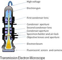
An electron microscope is a microscope that uses a beam of accelerated electrons as a source of illumination. As the wavelength of an electron can be up to 100, 000 times shorter than that of visible light photons, electron microscopes have a resolving power than light microscopes and can reveal the structure of smaller objects. Electron microscopes have electron optical lens systems that are analogous to the glass lenses of an optical light microscope.The similarity of scanning electron microscope (SEM) and transmission electron microscope (TEM) is are both structures smaller than 0.2 micrometer. The differences between of SEM and TEM are SEM produces a realistics 3D image of specimens's surface features but produces 2D image. The magnification of SEM is 1000X to 10,000X but in TEM are 10,000X to 100,000X. The resolving power for SEM is 20 nm but in TEM is 2.5 nm. SEM is to study the surface features of cells and viruses but TEM is to examine viruses or the internal ultrastructure in thin sections of cell. Electron Cryotomography is rapid technique provides way to preserve native state of structures examined in vacuum. Images recorded from many different directions to creates 3D structures.

Scanning Electron Microscope

view under electron microscope
Preparations of specimens for light microscopy
Preparations of specimens for light microscopy are wet mount and smears. Smears-steps involve are preparing smears, fixation, and staining.Fixation is a preserves internal and external structures and fixed them in position. Stain is a salt composed of a positive and negative ion, one of which is colourful this will called the chromophore. Stain make internal and external structures of cell more visible by increasing contrast with background. There are three types of staining which are simple staining, differential staining and special staining. In differential staining have gram staining and acid-fast staining. In special staining have negative staining, endospore staining and flagella staining.

gram stain

acid-fast staining

negative staining

endospore staining

flagella staining
Classification of organisms
Taxonomy is a arrangement into groups based on mutual similarities. There are three interrelated parts in the taxonomy which classification, identification and nomenclature. Classification is the arrangement of organisms into groups or taxon based on mutual similarity or evolutionary relatedness. Identification is the process of discovering and recording the traits of organisms. Nomenclature is a assignment of names to taxonomic groups in agreement with published rules.Strain is a subgroup of species with one or more characteristics that distinguish it from other subgroups of the same species. Different ways to describe strains within a species which are biovars, morphovars and serovars. Phylogenetic classifications is a natural systems based on evolutionary relationships and direct comparison of genetic material and gene products.
Methods of classifying and identifying microorganisms
Methods of classifying and identifying microorganisms are morphological characteristics, differential staining, biochemical tests, serology, phage typing, fatty acid profiles, DNA base composition,DNA fingerprinting, nucleic acid hybridization. Methods use to classify and identify microorganisms after various analyses which are dichotomous keys and cladograms.


No comments:
Post a Comment