Wednesday, 8 November 2017

Mircobiology Week 8 and experiments

        Assalamualaikum and Good afternoon everyone.... Hw r u guys? I hope you all fine. 😊😊😊This is my eighth week of my microbiology class and my basic techniques of microbiology class. In Basic technique of microbiology class, we did experiment 11 which is differential staining for visualization of bacterial cell structures. On wednesday, we all had our hand's on test which is practical test about streak plate isolation and Gram staining. In microbiology, Dr Wan asked us to make some questions about our this week microbiology topic which is eukaryotic cells by group and other every groups must answer the questions. On friday, Dr Wan asked us to chose one organelle and related organelles with algae, protozoa and fungi. Then, Dr Wan asked us to send the link of journals that we found.πŸ˜‰πŸ˜‰πŸ˜‰

Basic techniques of microbiology
  This week for a change Dr Fairolniza is our lecturer until the our last week of basic techniques of microbiology class. My first impression about Dr Fairol is strict. But is actually opposite to my impression, Dr Fairol is so friendly, nice and also beautiful like Dr Adelene. Dr Fairol give some briefing and explanation about the experiment 11. We did our experiment 11 by our ownself without teaching by Encik Hussain and Encik Zainuddin. On wednesday, as usual we observe the slide we are smear and stain. And also we did our hand's on test. Hand's on test like the demo gave us one unknown bacterial cultural and we have to isolate by using streak plate method and staining using Gram staining method. After that, we have to observe the slide under microscope and write the observation in the paper that provided to us. Lastly, after we finished observing our experiments, our demo gave some examples how to write the references for lab reports.

EXPERIMENT 11: Differential staining for visualization of bacterial cell structures

Part A: Spore stain

      Certain bacterial species, most commonly gram-positive bacilli such as those of the genera Bacillus and Clostridium, undergo a complex developmental cycle that produces a resting endospore when faced with environmental adversity. The process of sporulation allows the bacteria to survive in harsh environmental conditions such as low nutrients, high temperatures, UV radiation, acids and toxic chemicals. If conditions improve, the spore may germinate to form a new vegetative cell and growth will resume. These cells have the capacity to undergo sporogenesis and give rise to a new intracellular structure called the endospore, which is surrounded by imprevious layers called spore coats. Endospores are very dehydrated structures that are not metabolically active. They possess a protein coat, called an exosporium, that forms a barrier around the spore. Since endospores are not easily destroyed by heat or chemicals, they define the conditions necessary to sterility. For example, to destroy endospores by heating, they must be exposed for 15-20 minutes to steam under pressure, which generates temperatures of 121° C. As conditions continue to worsen, the endospore is released from the degenerating vegetative cell and becomes an independent cell called a free spore. With the return of favourable environmental conditions, the free spore may revert to a metabolically active and less resistant vegetative cell through germination. 

Primary stain
Malachite green used as the primary stain. Malachite green unlike most vegetative cell types that stain by common procedures, the free spores, because of its impervious coats, will not accept the primary stain easily. For further penetration, the application of heat is required. After the primary stain is applied and the smear is heated, both the vegetative cell and spore will appear green.

Decolorizing Agent
Water is used as the decolorizing agent. Once the spore accepts the malachite green, it cannot be decolorized by tap water, which removes only the excess primary stain The spore remains green. On the outer hand, the stain does not demostrate a strong affinity for vegetative cell components; the water removes it, and these cells will be colorless.

Counterstain 
Safranin is used as the counterstain. This contrasting red stain is used as the second reagent to color the decolorized vegetative cells, which will absorb the counterstain and appear red. The spores retain the green of the primary stain. 

Part B: Capsule Stain


          For capsule staining, the capsule stain employs an acidic stain and a basic stain to detect capsule production. A capsule is a gelatinous outer layer that is secreted by the cell and that surrounds and adheres to the cell wall. Capsules are formed by organisms such as Klebsiella pneumoniae . Most capsules are composed of polysaccharides, but some are composed of polypeptides. The capsule differs from the slime layer that most bacterial cells produce in that it is a thick, detectable, discrete layer outside the cell wall. Some capsules have well-defined boundaries, and some have fuzzy, trailing edges. Capsules protect bacteria from the phagocytic action of leukocytes and allow pathogens to invade the body. If a pathogen loses its ability to form capsules, it can become avirulent. Bacterial capsules are non-ionic, so neither acidic nor basic stains will adhere to their surfaces. Therefore, the best way to visualize them is to stain the background using an acidic stain and to stain the cell itself using a basic stain. We use India ink and Gram crystal violet. This leaves the capsule as a clear halo surrounding a purple cell in a field of black. Capsule staining is more difficult than other types of differential staining procedures because the capsular materials are water-soluble and may be dislodged and removed with vigorous washing. Smears should not be heated because the resultant cell shrinkage may create a clear zone around the organism that is an artifact that can be mistaken for the capsule.

Primary stain
        Crystal violet (1% aqueous) is used as primary stain. A violet stain is applied to a non-heat fixed smear. At this point, the cell and capsular material will take on the dark color. 

Decolorizing agent
      Copper sulfate (20%) is used as decolorizing agent because the capsule is nonionic, unlike the bacterial cell, the primary stain adheres to the capsules but does not bind to it. The copper sulfate washes the purple primary stain out of the capsular material without removing the stain bound to the cell wall. At the same time, the decolorized capsule will now appear blue in contrast to the deep purple color of the cell. 


Capsule staining of Enterobacter aerogenes using nigrosin 

Capsule staining of Klebsiella pneumoniae using nigrosin 

Spore staining of Bacillus cereus 

Capsule staining of Klebsiella pneumoniae using crystal violet

Capsule staining of Enterobacter aerogenes using crystal violet

Microbiology week 8 ( Eukaryotic cell) 

Endoplasmic reticulum


New findings challenge current view on origins of Parkinson's disease

    It was found that the bulk of the damage to neurons with damaged mitochondria stems from a related but different source -- the neighbouring maze-like endoplasmic reticulum (ER). The ER has the important job of folding proteins so that they can do the vast majority of work within cells. Misfolded proteins are recognized by the cell as being dangerous. Cells halt protein production if there are too many of these harmful proteins present. While this system is protective, it also stalls the manufacture of vital proteins, and this eventually results in the death of neurons. To find out if ER stress might be at play in Parkinson's, a team led by Dr Miguel Martins analyzed fruit flies with mutant forms of the pink1 or parkin genes. Mutant forms of pink1 and parkin are already known to starve neurons from energy by preventing the disposal of defective mitochondria. These genes are also mutated in humans and result in hereditary versions of the disease. Much like Parkinson's patients, flies with either mutation move more slowly and have weakened muscles. The insects struggle to fly and they lose dopaminergic neurons in their brains -- a classic feature of Parkinson's. Compared to normal flies, Miguel's team found that the mutants experienced large amounts of ER stress. The mutant flies did not manufacture proteins as quickly as the non-mutants. They also had elevated levels of the protein-folding molecule BiP, a telltale sign of stress.


Lysosomes

Macrophage Lysosome Damage Crucially Contributes to Fungal Virulence

     Upon ingestion by macrophages, Cryptococcus neoformans can survive and replicate intracellularly unless the macrophages become classically activated. The mechanism enabling intracellular replication is not fully understood; neither are the mechanisms that allow classical activation to counteract replication. C. neoformans–induced lysosome damage was observed in infected murine bone marrow–derived macrophages, increased with time, and required yeast viability. To demonstrate lysosome damage in the infected host, we developed a novel flow cytometric method for measuring lysosome damage. Increased lysosome damage was found in C. neoformans–containing lung cells compared with C. neoformans–free cells. Among C. neoformans–containing myeloid cells, recently recruited cells displayed lower damage than resident cells, consistent with the protective role of recruited macrophages. The magnitude of lysosome damage correlated with increased C. neoformans replication. Experimental induction of lysosome damage increased C. neoformans replication. Activation of macrophages with IFN-g abolished macrophage lysosome damage and enabled increased killing of C. neoformans. We conclude that induction of lysosome damage is an important C. neoformans survival strategy and that classical activation of host macrophages counters replication by preventing damage. Thus, therapeutic strategies that decrease lysosomal damage, or increase resistance to such damage, could be valuable in treating cryptococcal infections.

Peroxisome

Antifungal activity of Saccharomyces cerevisiae peroxisomal 3-ketoacyl-CoA thiolase

       Peroxisomes play an important role in cellular defense systems and generate secondary messengers for cellular communication. Saccharomyces cerevisiae containing oleate-induced peroxisomes were subjected to buffer-soluble extraction and two chromatographicprocedures, and a protein with antifungal activity was isolated. The results of MALDI-TOF analysis identified the isolated protein as peroxisomal 3-ketoacyl-CoA thiolase (ScFox3). Purified yeast ScFox3 exhibited thiolase activity that catalyzed the thiolytic cleavage of 3-ketoacyl-CoA to acetyl-CoA and acylCoA. ScFox3 protein inhibited various pathogenic fungal strains, with the exception of Aspergillus flavus. Using ScFox3-GFP and PTS2 signal-truncated ScFox3M-GFP, we showed that only ScFox3-GFP, with an intact PTS2 peroxisome signal sequence, was able to translocate into peroxisomes. Yeast ScFox3 is a natural antifungal agent found in peroxisomes.

Vacuoles

Light Shielding in Blue-Green Algae

       Gas vacuoles are small, cylindrical, gas-filled vesicles which occur in certain procaryotic cells. They are found in numerous blue-green algae, many photosynthetic bacteria, some halophilic bacteria, and some planktonic freshwater bacteria. Because gas-vacuolate blue-green algae are often observed floating at the surface of water where high light intensity may damage cells, Lemmerman first suggested that gas vacuoles may function as light-shielding organelles. Recently, it has been suggested that such light shielding (if it occurred) might be due to the optical properties and the intracellular distribution of the gas vacuoles. Being gas filled, these vacuoles have a refractive index much lower than that of the cytoplasm which surrounds them, and therefore they scatter light. A suspension of gas vacuolate cells becomes visibly less milky and more transparent when the gas vacuoles are collapsed. Similarly, when a milky-white, opalescent suspension of isolated gas vacuoles is subjected to sudden pressure, a completely transparent suspension results. These changes are due to a decrease in light scattering when gas vacuoles collapse. But, although light scattering by gas vacuoles could protect cells by scattering away a large portion of the incident radiation, it could also lead to increased rather than decreased irradiation of some cellular components if the light were back-scattered into these components. Whether gas vacuoles function as light shields would depend upon the distribution of the gas vacuoles inside the cells.

Chloroplast

Toward mosquito control with a green alga: Expression of Cry toxins of Bacillus thuringiensis subsp. israelensis (Bti) in the chloroplast of Chlamydomonas.

        We are developing Chlamydomonas strains that can be used for safe and sustainable control of mosquitoes, because they produce proteins from Bacillus thuringiensis subsp. israelensis (Bti) in the chloroplast. Chlamydomonas has a number of advantages for this approach, including genetic controls that are not generally available with industrial algae. The Bti toxin has been used for mosquito control for > 30 years and does not engender resistance; it contains three Cry proteins, Cry4Aa (135 kDa), Cry4Ba (128 kDa) and Cry11Aa (72 kDa), and Cyt1Aa (25 kDa). To express the Cry proteins in the chloroplast, the three genes were resynthesized and cry4Aa was truncated to the first 700 amino acids (cry4Aa700 ); also, since they can be toxic to host cells, the inducible Cyc6:Nac2-psbD expression system was used. Western blots of total protein from the chloroplast transformants showed accumulation of the intact polypeptides, and the relative expression level was Cry11Aa > Cry4Aa700 > Cry4Ba. Quantitative western blots with purified Cry11Aa as a standard showed that Cry11Aa accumulated to 0.35% of total cell protein. Live cell bioassays in dH20 demonstrated toxicity of the cry4Aa700 and cry11Aa transformants to larvae of Aedes aegypti and Culex quinquefasciatus. These results demonstrate that the Cry proteins that are most toxic to Aedes and Culex mosquitoes, Cry4Aa and Cry11Aa, can be successfully expressed in the chloroplast of Chlamydomonas.

Mitochondria

Hydrogen Production. Green Algae as a Source of Energy

       Hydrogen gas is thought to be the ideal fuel for a world in which air pollution has been alleviated, global warming has been arrested, and the environment has been protected in an economically sustainable manner. Hydrogen and electricity could team to provide attractive options in transportation and power generation. Interconversion between these two forms of energy suggests on-site utilization of hydrogen to generate electricity, with the electrical power grid serving in energy transportation, distribution utilization, and hydrogen regeneration as needed. A challenging problem in establishing H2 as a source of energy for the future is the renewable and environmentally friendly generation of large quantities of H2 gas. Thus, processes that are presently conceptual in nature, or at a developmental stage in the laboratory, need to be encouraged, tested for feasibility, and otherwise applied toward commercialization.

Nuclues

Regulation of eukaryotic DNA replication and nuclear structure

     In eukaryote, nuclear structure is a key component for the functions of eukaryotic cells. More and  more evidences show that the nuclear structure plays important role in regulating DNA replication. The nuclear structure provides a physical barrier for the replication licensing, participates in the decision where DNA replication initiates, and organizes replication proteins as replication factory for DNA replication. Through these works, new concepts on the regulation of DNA replication have emerged, which will be discussed in this minireview. Regulatory mechanisms for DNA replication are central to the control of the cell-cycle in eukaryotic cells. Recently, considerable progress has been made in our understanding of the relationship between regulation of eukaryotic DNA replication and nuclear structure. This review will briefly outline the progress and discuss some new concepts
appearing from the studies.

Image result for eukaryotic cells         Image result for eukaryotic cells


Thursday, 2 November 2017

Microbiology Week 7 and Experiments

      Assalmualaikum and good afternoon everyone. Hw r u? I hope you all fine as well. 😁😁😁 This week is my seventh week of microbiology class and this week I like to combine my post of microbiology and my basic technique of microbiology because they are interconnected with each other. In my basic technique class we did experiments 9 which is Gram staining and experiment 10 which is Acid fast staining. In my microbiology class, on wednesday we all having our suspend quiz on that but it is not individual quiz, it is team quiz about intracellular structure of prokaryotes  πŸ˜”πŸ˜”πŸ˜”and today we are going to study about intracellular structure of prokaryotes.


Basic techniques of microbiology 

EXPERIMENT 9 : Gram staining and EXPERIMENT 10: Acid fast staining
   
    After we enter the class, Dr Adelene talked about our test and what the mistake we did in our test. Then, Dr Adelene give some briefing and introduction about the experiment 9 and 10. For a change, this week Encik Zainuddin didn't give some demo how to do this two experiments. We did the experiments by our ownself. And after we finished our experiments, my demo was talked about the mistakes of the previous lab report. Half of my classmate did wrong in experiment A which is about measurement of microorganisms and again she taught us how to calculate it. So now I'm going to share the information about what I got learned in this week about experiment 9 and 10. 
                                                                   Before staining
After staining 


Experiemnt 9 : Gram staining

 The Gram stain is the most important stain procedure in microbiology. It is used to differentiate between gram positive organisms and gram negative organisms. Hence, it is a differential staining.  Differential staining requires the use of at least four chemical reagents that are applied sequentially to a heat-fixed smear. The first reagent is called primary stain is to impart it's color to all cells. The second stain is a mordant used to intensify the color of the primary stain. Based on the chemical composition of cellular components, the decolorizing agent may or may not remove the primary stain from the entire cell or only from certain cell structures. The final reagent is the counterstain, has a contrasting color to that of the primary stain. Gram stain reaction is based on the difference in the chemical composition of bacterial cell walls. Gram positive cell have thick layer of peptidoglycan but gram negative have thin layer of peptidoglycan and surrounded by outer lipid-containing layers. Peptidoglycan is mainly a polysaccharides composed of two chemical subunits found only in the bacterial cell wall. These subunits are N-acetylglucosamine and N-acetylmuramic acid.

Primary stain
    The primary stain is used in Gram staining to detect the Gram positive bacteria. The primary stain is a crystal violet color. When a bacteria is Gram negative, it loses the primary stain and takes on the counterstain of safranin. The violet stain is used first and stains all cells purple.

Mordant
    A mordant is a chemical used to hold down molecules of a stain onto a microorganism. Classically defined, mordants are usually ions such as metal ions or halide ions, but can be any molecule that serves the purpose of holding down a dye. However, a molecule called phenol is a non-ionic mordant that is discussed below. Some mordants bind both the dye and proteins on the microorganism. Most mordants are ions because the electrical charge on the ion attracts the electrical charge on a chemical dye. Thus, when the ion binds the dye, they form a large complex that precipitates -- meaning they become a solid and are no longer dissolved in the solution. Mordants hold down, or weigh down, the dye so it does not wash away during the remainder of the staining procedure. Washing is done so that only the true staining regions are visualized. At this point, all cells appear purple-black.

Decolorizing agent
    Alcohol or acetone dissolves the lipid outer membrane of Gram negative bacteria, thus leaving the peptidoglycan layer exposed and increases the porosity of the cell wall. The CV-I complex is then washed away from the thin peptidoglycan layer, leaving Gram negative bacteria colorless. On the other hand, alcohol has a dehydrating effect on the cell walls of Gram positive bacteria which causes the pores of the cell wall   to shrink. The CV-I complex gets tightly bound into the multi-layered, highly cross-linked Gram positive cell wall thus staining the cells purple. The decolorization step must be performed carefully, otherwise over-decolorization may occur. This step is critical and must be timed correctly otherwise the crystal violet stain will be removed from the Gram-positive cells. If the decolorizing agent is applied on the cell for  too long time , the Gram-positive organisms to appear Gram-negative. Under-decolorization occurs when the alcohol is not left on long enough to wash out the CV-I complex from the Gram-negative cells, resulting in Gram-negative bacteria to appear Gram-positive. Thus, the tightly bound primary stain complex is difficult to remove, and the cells remain purple.

Counterstain
    The decolorized Gram negative cells can  be rendered visible with a suitable counterstain, which is usually positively charged safranin, which stains them pink. Pink colour which adheres to the Gram positive bacteria is masked by the purple of the crystal violet.

Experiment 10: Acid fast staining

     The acid-fast stain is a differential stain used to identify acid-fast organisms such as members of the genus Mycobacterium. Acid-fast organisms are characterized by wax-like, nearly impermeable cell walls; they contain mycolic acid and large amounts of fatty acids, waxes, and complex lipids.  Acid-fast organisms are highly resistant to disinfectants and dry conditions. Because the cell wall is so resistant to most compounds, acid-fast organisms require a special staining technique. The primary stain used in acid-fast staining, carbolfuchsin, is lipid-soluble and contains phenol, which helps the stain penetrate the cell wall. This is further assisted by the addition of heat. The smear is then rinsed with a very strong decolorizer, which strips the stain from all non-acid-fast cells but does not permeate the cell wall of acid-fast organisms. The decolorized non-acid-fast cells then take up the counterstain. In our lab, we use the Ziehl-Neelsen method but not the Kinyoun method.

Primary stain
    Carbol fuchsin is a dark red stain in 5% phenol that is soluble in the lipoidal materials that constitute most of the mycobacterial cell wall, does penetrate these bacteria and its retained. Penetration is further enhanced by the application of heat, which drives the carbol fuchsin through the lipoidal wall and into the cytoplasm. This appllication of heat is used in the Ziehl-Neelsen method. The Kinyoun method, a modification of the Ziehl-Neelsen method, a modification of the Ziehl-Neelsen method, circumvents the use of heat by addition of a wetting agent to this stain, which reduces surface tension between the cell wall of the mycobacteria and the stain. Following application of the primary stain, all cells will appear red.

Decolorizing agent
      On application of acid alcohol (3% HCL + 95% Ethanol), the acid-fast cell will be resistant to decolorization, since the primary stain is more soluble in the cellular waxes than the decolouring reagent. In this event, the primary stain is retained and the mycobacteria will stay red. This is not the case with the nonacid-fast organisms that lacks the cellular waxes in their cell wall. The primary stain is more easily removed during decolorization, leaving these cells colourless or unstained.

Counterstain
Methylene blue is used as the final reagent to stain previously depolarized cell. As only nonacid-fast cells undergo decolourization, they may now absorb the counter stain and take on its blue color, while acid-fast cells retain the red color of the primary stain.
                                                 
                                  Staphylococcus aureus in Gram stain
                                                               

  Mixture of Mycobacterium smegmatis and Staphylococcus aureus in Acid fast stain

                                 Mycobacterium smegmatis in Acid fast stain

Mixture of Escherichia coli and Bacillus cereus in Gram stain


Microbiology Week 7 ( Intracellular structures of prokaryotes)
   
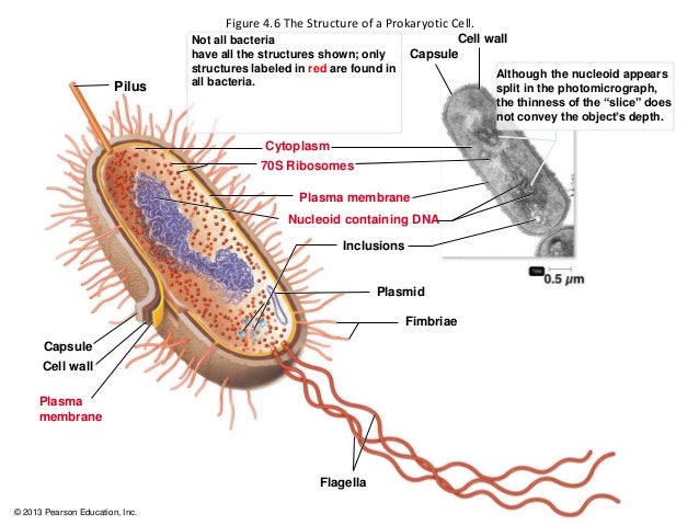
      The intracellular structures of prokaryotes which are plasma membrane, cytoplasm, the nuclear area, ribosomes, inclusions and endospore. Plasma membrane structure is phospholipid bilayer with proteins embedded in and attached to inner and outer surfaces. The function of the plasma membrane is selectively permeable barrier which is phospholipids are liquid at body temperature functions as a Fluid Mosaic, synthesizes cell wall components, assists in DNA replication, carries on respiration and captures energy as ATP. Destruction of the plasma membrane is disinfectants which is alcohol and quaternary ammonium compunds cause leakage of intracellular contents. The movement of materials across membranes is passive processes and active processes. Passive processes is molecules cross the membrane from an area of high concentration to an area of low concentration and the concentration gradient not energy dependent. Passive processes can classify into three processes which is simple diffusion, facilitated diffusion and osmosis. Simple diffusion is movement of molecules or ions from high to low concentration until equilibrium. Facilitated diffusion is a substances are moved by transporter proteins from high to low concentration and  needs a carrier proteins as transporters. Osmosis is movement of water molecule from high to low concentration. Active process is energy dependent system and ATP or proton motive force used. Active process can classify into two process which is active transport and group translocation. Active transport is substances are moved by transporter proteins from low to high concentration and cell has to expend energy for this to happen. Group translocation is molecules are chemically modified during passage across cytoplasmic membrane and energy is expended.
       Cytoplasm contains proteins (enzymes), carbohydrates, lipids, inorganic ions and many low molecular weight compounds. The major structures in the prokaryotic cytoplasm are DNA, ribosomes and inclusions. In intracytoplasmic membranes, plasma membrane infoldings is observed in many photosynthetic bacteria and observed in many bacteria with high respiratory activity and Anammoxosome in Planctomycetes is the organelle is site of anaerobic ammonia oxidation.
       The nuclear area is single long circular molecule of double-stranded DNA (bacterial chromosome). Bacterial chromosome do not include histones and are not sirrounded by nuclear envelope. In addition to the bacterial chromosome, bacteria often contain small circular, double-stranded DNA molecules called plasmid. Plasmid is a small circular, and double-stranded DNA. Extrachromosomal genetic elements not connected to bacterial chromosome and replicate independently of chromosomal DNA. Plasmid do not contain genetic material essential for growth. Plasmid is also contains features that enhance survivability examples of gene for drug resistance and it is also transferable from one bacterial to another.
      The function of the ribosome is sites of protein synthesis. It is consists of 70S ribosome. Ribosome has two subunit (small subunit-30S subunit and large subunit- 50S subunit). S refer to Svedberg unit. Each subunit consists of protein and RNA called ribosomal RNA or rRNA. Ribosome can be inhibited by certain antibiotics. Ribosome's cells that have high rates of protein synthesis, have a large number of ribosomes. Several antibiotics work by inhibiting protein synthesis on prokaryotic ribosomes which is streptomycin and gentamicin attach to the 30S subunit and erythromycin and chloramphenical attach to the 50S subunit.
    Inclusions is reserve deposits and can serve as a basis of identification. The type of inclusions are metachromatic granules, polysaccharide granules, lipid inclusions, sulfur granules, carboxysomes, magnetosomes and gas vacoule. Metachromatic granules is the large inclusion and stain red with certain blue dyes such as methylene blue. Metachromatic granules collectively known volutin is the inorganic phosphate and the phosphate used in the synthesis of ATP. Polysaccharide granules consist of glycogen and starch. Differentiated of polysaccharide granules by using iodine, reddish brown indicates glycogen granules and blue indicates starch granules. Lipid granules is storage material which is polymer poly-Ξ²-hydroxybutyric acid and revealed by Sudan dyes (fat-soluble). Sulfur granules is serves as an energy reserve. Derive energy by oxidising sulfur and sulfur containing compunds. Carboxysomes is contain the enzyme ribulose 1,5-diphosphate carboxylase and used for carbon dioxide fixation during phtosynthesis. Magnetosomes is a iron oxide that act like magnets and for downward movement until reaching suitable attachment site. Function of magnetosomes is to protect the cell against hydrogen peroxide accumulation. Gas vesicles is a hallow cylinder covered by proteins and collectively called gas vacuole. Gas vesicle appeared bright, refractile areas with an irregular outline in the phase microscope and consists of thin membrane surrounding a hallow space. Function of gas vesicles is to provide buoyancy for aquatic prokaryotes to receive sufficient amounts of oxygen, light and nutrients.
      Endospore is a resting structures formed by some bacteria for survival during adverse environmental conditions and germination results in leaving the dormant stage and once again becoming a typical, multiplying cell (vegetative cell). Genus Bacillus and Clostridium are two common disease causing bacteria that produce endospores as needed. Endospores cannot be destroyed easily, even by harsh chemicals, are formed internal to the bacterial cell membrane, highly resistant differentiated bacterial cell, and enable the organism to endure extreme environmental conditions. Endospore is a formation leads to a highly dehydrated structure thick walls and additional layers and contain essential macromolecules and a variety of substances absent from vegetative cells. Endospore called because the spore is formed within the cell. Endospores's spores are very impermeable to dye. However, they can be stained with special dye, Malachite green. Endospore is also small acid-soluble proteins to protect DNA from UV radiation, desiccation, and dry heat and serve as a carbon and energy source during germination. Endospore can remain dormant indefinitely but germinate quickly when the appropriate trigger is applied. Endospores and Infectious disease, although harmless themselves until they germinate, they are involved in the transmission of some disease to humans. Infections transmitted to humans by endospores included Bacillus anthracis, Clostridium tetani, Clostridium botulinum, and Clostridium perfringens.  
   Image result for intracellular structures of prokaryotes


Monday, 30 October 2017

Microbiology Week 6

    Assalamualaikum and good evening to everyone that read my post.... Hw r u everyone? I hope everyone will fine. 😁😁😁 This week is my sixth week of microbiology class and my test 1 is also in this week. πŸ˜‘πŸ˜‘πŸ˜‘ Everyweek have two microbiology but this week only one microbiology class because in another class, we all did out test 1 of microbiology. The test quiet difficult not all questions are difficult. There are some of the questions are easy. Usually on wednesday is my  microbiology class, but for change my microbiology class on tuesday. On tuesday, we learned about introduction to prokaryotes. The class start at 12 to 1pm. That time usually I'm so sleepy 😴😴😴 and I undrrstand a bit about the topic. Dr Wan also quiet fast when taught us that day. So now, I'm going to share about what I learned for this week.

    The prokaryotes are a group of organisms that lack a cell nucleus or any other membrane-bound organelles. Most are unicellular, but a few prokaryotes such as myxobacteria have multicellular stages in their life cycles. The size of prokaryotes is 0.2 to 2.0ΞΌm in diameter and 2 to 8ΞΌm in length. There are three basic shapes which are coccus, bacillus and spiral. Examples of cocci are diplococci, staphylococci, and streptococci. Examples of bacilli are diplobacilli and streptobacilli. Examples of spiral are vibrio, spirillum and spirochetes. Diplococci is in pairs, streptococci is in chain, staphylococci is grape-like cluster, tetrads is four cocci in a square and sarcinae in cubic configuration of eight cocci. The structures external to the cell wall is glycocalyx, flagella, axial filaments and fimbriae and pili. Glycocalyx is substance that surround cell and it is made inside the cell and excreted to the cell surface. The function of glycocalyx is protection from phygocytosis, attachment to various surfaces, source of nutrients and protect a cell against dehydration. Flagella is threadlike, locomotor appendages extending outward from plasma membrane and cell wall. Th e function of flagella is motility and swarming behavior, attachment to surfaces and may be virulence factors. The axial filaments is bundle of fibrils that arise at the ends of the cell beneath the outer sheath. Fimbriae and pili is hairlike appendages that are shorter, straighter and thinner than flagella. It consist of a protein called pilin arranged helically around a central core.

    The cell wall surrounds the cytoplasmic membrane and not a regulatory structure like cytoplasmic membrane. Composition and characteristics of cell wall is composed of a macromolecular network called peptidoglycan. Peptidoglycan consists of a repeating disaccharide. The disaccharide portion is made up of monosaccharieds called N-acetylglucosamine (NAG) and N-acetylmuramic acid (NAM). Alternating NAM and NAG molecules are linked in row by glycosidic bonds and adjacent rows are linked by peptide bonds. Functions of cell wall is prevent bacterial cell from rupturing when the water pressure inside the cell is greater than that outside the cell, contributes to pathogenicity, maintain characteristics shape, provides a rigid platform and counters the effect of osmotic pressure. Cell wall have two major types of walls which are Gram-positive and Gram-negative. Gram positive cell walls consists of many layers of peptidoglycan. Periplasmic space of Gram positive bacteria lies between plasma membrane and cell wall and is smaller than that of Gram- negative bacteria. Teichoic acid is primarily of an alcohol and phosphate, negatively charged, provide much of the wall' s antigenic specificity and cell walls do not degrade as easily. Two classes of teichoic acids which are lipoteichoic acid and wall teichoic acid. Gram negative cell wall consists of one or very few layers of peptidoglycan . The peptidoglycan is bonded to lipoprotein and does not contain teichoic acid. Periplasmic contains a high concentration of degrading enzymes and large number of transport proteins. Gram-negative cell wall consist of outer membrane is outer membrane consists of lipoproteins, lipopolysaccharides, phospholipids and porins, strong negative charge, and provide a barrier to certain antibiotic, lysozyme, detergent, heavy metals, bile salts and certain dyes.

     Atypical cell walls is mycoplasma, chlamydiaceae and archaea. Mycoplasma cells have no walls or have very little wall material. It's membrane contain sterols, which impart rigidity to the membrane and can pass through most bacterial filters. Plasma membranes are unique in having lipids called sterols which protect them from osmotic lyses. Chlamydiaceae have two membrane, some genes for peptidoglycan synthesis found in genome and obligate intracellular parasites. Archaea are lack of peptidoglycan in their cell walls. Some species have cell wall consisting of polysaccharide, glycoprotein or protein but not peptidoglycan. However, contain a substance similar to peptidoglycan called pseupeptidoglycan. Most common wall type is paracrystalline surface layer (S-layer) made of protein or glycoprotein with hexagonal symmetry. S-layer surface, or outermost layer forming "lipidless membrane", found on Archaea a few in Gram-positive and Gram-negative, composed  of repeating subunits of protein or glycoprotein, protect against osmotic stress, pH and enzyme and aid in attachment and inhibit phagocytosis. Archaea are naturally resistant to lusozyme and penicillin.


Image result for prokaryote               Image result for prokaryote


Image result for mycoplasma       

Friday, 13 October 2017

Microbiology Week 5

        Assalamualaikum and good morning everyone... How are you all ? πŸ˜ƒπŸ˜ƒπŸ˜ƒ This week is my fifth week of microbiology class huhu.... But actually I not yet adapt the environment in University Putra Malaysia as a degree student of microbiology. This week many things happened in my microbiology class. Firstly, For two days continuously Dr Wan gave some " tazkirah" which means advice to us because in class we are like not more curious to know about the microbes and all the things related to microbes. And also we even didn't look at what my friends posted in Open learning MOOC about the interesting things which are about Helicobacter pylori and Microbial interactions in termite guts. So, Dr Wan asked to make a group and each group have to choose one bacteria and we have to present it in three minutes. For my group, we were chose Magnetotatic bacteria. The Magnetotatic bacteria are quiet interesting because it is about magnetic. Others group also were chose about Mycobacterium tuberculosis, Serratia marcescens, Propiniobacterium acnes, Alcanivorax borkumensis and Pseudomonas aeruginosa. We were actually presented about our bacteria on friday. Each group must present 3 minutes, but some group presented more than 3 minutes because they want us to be more clear about what they are presented. Everyone were presented in our microbiology class for first time.   In my group , we were separated our work like one persons find about the history of that bacteria another about the characteristics of bacteria that my group chose. It is like team work and it make more closer to each other. So now I'm going to share about what I had studied for this week.

 Methods of classifying  and identifying microorganisms

        Morphological characteristics is easy to study and analaysed. Characteristics studied include cell type , shape and size, cellular grouping , external structures and internal structures. Differential staining which are Gram stain is classifies into gram positive or negative, Acid-fast stain is only stain bacteria with waxy material in their cell walls, Negative staining for capsule is use India ink to provide a contrasting background, then stain with simple stain and capsules to not accept simple stain, thus appear as halos surrounding bacterial cell, Endospore staining is appear green within pink cells and Flagella staining are too thin and use a mordant and stain carbolfuchsin to coat the flagella until they are thick enough to be seen. Biochemical test to verify its metabolic activity examples are Phenol Red broth, Gelatin test, Lipase test, Starch hydrolysis, Motility test and Catalase test. Serology is the science that studies serum and immune responses that are evident in serum and the background are Bacteria (antigen) enter a host, antibody will be produced by the host. The antibody will combine with the antigen ( bacteria) and this precipitates the antigen. Two types of serology which are Enzyme-linked immunosorbent assay (ELISA) is fast and utilised a computer scanner to read result and performed in microtiter plate and Western blotting is protein from an unknown bacterium or virus are separated by electrophoresis and transferred to a nitrocellulose filter by blotting.  Phage typing is to determine which phage a bacterium is susceptible to bacteriophage cause lysis of bacteria that they infect and infect only particular species or even strains. Fatty acid profiles is bacteria synthesise a wide variety of fatty acids these fatty acids are constant for a particular species. DNA Base Composition is the base composition of a single species is a fixed property that can be used to reveal the degree of species relatedness. DNA Fingerprinting is use of restriction enzyme to produce banding pattern. Nucleic Acid Hybridization can be used to determine extent of similarity based on degree of reunion. Techniques that apply the principle of nucleic acid hybridization are sounthern blotting, DNA chips, Ribotyping and Ribosomal RNA Sequencing and Fluorescent In Situ Hybridization. Methods use to classify and identify microorganisms after various analyses are Dichotomous Keys used for identification based on successive questions and Cladograms is show evolutionary relationships among organisms and constructed based on rRNA sequences with the aid of software.

Mycobacterium tuberculosis

Mycobacterium tuberculosis discovered by Robert Koch in 1882. Mycobacterium tuberculosis is a non motile, acid-fast, obligate aerobe. The bacilli are 2-4 micrometer in length and have a very slow generation of time between 15 to 20 hours. The cell wall of the Mycobaterium is unique in that it is composed mainly of acidic waxes, specifically mycolic acids. Mycobacterium tuberculosis is usually resistant to drying and chemicals, contributing to the ease with which it is transmitted. Mycobacterium tuberculosis is ropelike structure with peptidoglycan and found abundantly in soil. The tuberculin skin test currently used to diagnose infection with Mycobacterium tuberculosis has poor diagnostic value, especially in geographic areas where the prevalence of tuberculosis is low or where the environment burden of saprophytic, nontuberculous mycobacteria is high.


Alcanivorax borkumensis

Alcanivorax borkumensis is an alkane-degrading marine bacterium which naturally propagates and becomes predominant in crude-oil-containing seawater when nitrogen and phosphorus nutrients are supplemented. They are currently thought to be the world's most important oil-degrading organisms. This bacteria is useful in treating oil spillage because it is an oil-eating bacteria. A borkumensis is a rod-shaped bacterium without flagella that obtains its energy primarily from consuming alkanes. It is aerobic meaning it uses oxygen to gain energy and it is halophilic, meaning it tends to live in environments that contain salt, such as salty ocean water. It is also Gram-negative which essentially means it has a relatively thin cell wall. It is also non-motile, however other organisms that appear to be in the same genus are motile through flagella.


Propionibacterium acnes

 Propionibacterium acnes is the relatively slow-growing, typically aerotolerant anaerobic, Gram-positive bacterium (rod) linked to the skin condition of acne. P. acnes bacteria live deep within follicles and pores, away from the surface of the skin. In these follicles, P. acnes bacteria use sebum, cellular debris and metabolic byproducts from the surrounding skin tissue as their primary sources of energy and nutrients. Elevated production of sebum by hyperactive sebaceous glands (sebaceous hyperplasia) or blockage of the follicle can cause P. acnes bacteria to grow and multiply. P. acnes bacteria secrete many proteins, including several digestive enzymes. These enzymes are involved in the digestion of sebum and the acquisition of other nutrients. They can also destabilize the layers of cells that form the walls of the follicle. The cellular damage, metabolic byproducts and bacterial debris produced by the rapid growth of P. acnes in follicles can trigger inflammation. This inflammation can lead to the symptoms associated with some common skin disorders, such as folliculitis and acne vulgaris. P. acnes bacteria are susceptible to a wide range of antimicrobial molecules, from both pharmaceutical and natural sources. Antibiotics are commonly used to treat infections caused by P. acnes. Acne vulgaris is the disease most commonly associated with P. acnes infection. The antibiotics most frequently used to treat acne vulgaris are erythromycin, clindamycin, doxycycline, and minocycline. Several other families of antibiotics are also active against P. acnes bacteria, including quinolones, cephalosporins, pleuromutilins, penicillins, and sulfonamides.


Thursday, 5 October 2017

Microbiology Week 4

   Assalamualaikum and good afternoon eveyone that reading my blog now.... πŸ˜ƒπŸ˜ƒπŸ˜ƒπŸ˜ƒ. This week is my forth week of microbiology class. I feel like it is my first class but actually my forth class with Dr Wan... You know time flying so fast. I would like to share you what I learned in the forth week of microbiology class with my spotting lecturer Dr Wan. Usually after my microbiology class, I feel so tired and sometimes I a bit stress because a lot of works and reseach have to do. That was as usual because I as a microbiology student I have to do more reasearch to know and find the types of microoorganisms. Like a scientist huhuhu 😍😍😍. I feel stress because Im not yet adapt the changes in university. I think it will takes a bit more time to adapt the changing because now only my enter to the degree. But for sure oneday i will change and become a good and profesional scientist .😎😎😎 So on wednesday, Dr Wan taught us about electron microscope and on friday we all had group discussion about the classification of organisms. Group discussion is like in one group we have to separate the topic among with our group members and understand about the topics. Then who are got the same topics to be discuss they are will combine and make a group. They discuss about what they are understand and do more research. Afterthat, only two people are in the group and the rest of all can go to another topic groups and the another group members will explain about what they are understand about their topics. The discussion a quiet fun and we all enjoyed but we does not have enough time to go to more groups but Dr Wan said we will study again in next week.This week for classifications of organisms are short details. After Dr Wan explain it then most probably next week I will share more about classifications of organisms. So, Im going to share the information about electron microscope.

              Electron Microscope

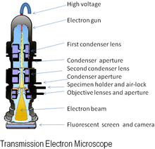
An electron microscope is a microscope that uses a beam of accelerated electrons as a source of illumination. As the wavelength of an electron can be up to 100, 000 times shorter than that of visible light photons, electron microscopes have a resolving power than light microscopes and can reveal the structure of smaller objects. Electron microscopes have electron optical lens systems that are analogous to the glass lenses of an optical light microscope.The similarity of scanning electron microscope (SEM) and transmission electron microscope (TEM) is are both structures smaller than 0.2 micrometer. The differences between of SEM and TEM are SEM produces a realistics 3D image of specimens's surface features but produces 2D image. The magnification of SEM is 1000X to 10,000X but in TEM are 10,000X to 100,000X. The resolving power for SEM is 20 nm but in TEM is 2.5 nm. SEM is to study the surface features of cells and viruses but TEM is to examine viruses or the internal ultrastructure in thin sections of cell. Electron Cryotomography is rapid technique provides way to preserve native state of structures examined in vacuum. Images recorded from many different directions to creates 3D structures.


Scanning Electron Microscope

view under electron microscope
         

Preparations of specimens for light microscopy 

Preparations of specimens for light microscopy are wet mount and smears. Smears-steps involve are preparing smears, fixation, and staining.Fixation is a preserves internal and external structures and fixed them in position. Stain is a salt composed of a positive and negative ion, one of which is colourful this will called the chromophore. Stain make internal and external structures of cell more visible by increasing contrast with background. There are three types of staining which are simple staining, differential staining and special staining. In differential staining have gram staining and acid-fast staining. In special staining have negative staining, endospore staining and flagella staining.

gram stain

acid-fast staining 

negative staining

endospore staining 

flagella staining 


Classification of organisms 

Taxonomy is a arrangement into groups based on mutual similarities. There are three interrelated parts in the taxonomy which classification, identification and nomenclature. Classification is the arrangement of organisms into groups or taxon based on mutual similarity or evolutionary relatedness. Identification is the process of discovering and recording the traits of organisms. Nomenclature is a assignment of names to taxonomic groups in agreement with published rules.Strain is a subgroup of species with one or more characteristics that distinguish it from other subgroups of the same species. Different ways to describe strains within a species which are biovars, morphovars and serovars. Phylogenetic classifications is a natural systems based on evolutionary relationships and direct comparison of genetic material and gene products.

Methods of classifying and identifying microorganisms

Methods of classifying and identifying microorganisms are morphological characteristics, differential staining, biochemical tests, serology, phage typing, fatty acid profiles, DNA base composition,DNA fingerprinting, nucleic acid hybridization. Methods use to classify and identify microorganisms after various analyses which are dichotomous keys and cladograms.


Microbiology Semester 2 Week 14

              Assalamualaikum and hi everyone... how are you all? I hope everyone will be fine as well... On Tuesday, before our class start...