Monday, 30 October 2017

Microbiology Week 6

    Assalamualaikum and good evening to everyone that read my post.... Hw r u everyone? I hope everyone will fine. 😁😁😁 This week is my sixth week of microbiology class and my test 1 is also in this week. πŸ˜‘πŸ˜‘πŸ˜‘ Everyweek have two microbiology but this week only one microbiology class because in another class, we all did out test 1 of microbiology. The test quiet difficult not all questions are difficult. There are some of the questions are easy. Usually on wednesday is my  microbiology class, but for change my microbiology class on tuesday. On tuesday, we learned about introduction to prokaryotes. The class start at 12 to 1pm. That time usually I'm so sleepy 😴😴😴 and I undrrstand a bit about the topic. Dr Wan also quiet fast when taught us that day. So now, I'm going to share about what I learned for this week.

    The prokaryotes are a group of organisms that lack a cell nucleus or any other membrane-bound organelles. Most are unicellular, but a few prokaryotes such as myxobacteria have multicellular stages in their life cycles. The size of prokaryotes is 0.2 to 2.0ΞΌm in diameter and 2 to 8ΞΌm in length. There are three basic shapes which are coccus, bacillus and spiral. Examples of cocci are diplococci, staphylococci, and streptococci. Examples of bacilli are diplobacilli and streptobacilli. Examples of spiral are vibrio, spirillum and spirochetes. Diplococci is in pairs, streptococci is in chain, staphylococci is grape-like cluster, tetrads is four cocci in a square and sarcinae in cubic configuration of eight cocci. The structures external to the cell wall is glycocalyx, flagella, axial filaments and fimbriae and pili. Glycocalyx is substance that surround cell and it is made inside the cell and excreted to the cell surface. The function of glycocalyx is protection from phygocytosis, attachment to various surfaces, source of nutrients and protect a cell against dehydration. Flagella is threadlike, locomotor appendages extending outward from plasma membrane and cell wall. Th e function of flagella is motility and swarming behavior, attachment to surfaces and may be virulence factors. The axial filaments is bundle of fibrils that arise at the ends of the cell beneath the outer sheath. Fimbriae and pili is hairlike appendages that are shorter, straighter and thinner than flagella. It consist of a protein called pilin arranged helically around a central core.

    The cell wall surrounds the cytoplasmic membrane and not a regulatory structure like cytoplasmic membrane. Composition and characteristics of cell wall is composed of a macromolecular network called peptidoglycan. Peptidoglycan consists of a repeating disaccharide. The disaccharide portion is made up of monosaccharieds called N-acetylglucosamine (NAG) and N-acetylmuramic acid (NAM). Alternating NAM and NAG molecules are linked in row by glycosidic bonds and adjacent rows are linked by peptide bonds. Functions of cell wall is prevent bacterial cell from rupturing when the water pressure inside the cell is greater than that outside the cell, contributes to pathogenicity, maintain characteristics shape, provides a rigid platform and counters the effect of osmotic pressure. Cell wall have two major types of walls which are Gram-positive and Gram-negative. Gram positive cell walls consists of many layers of peptidoglycan. Periplasmic space of Gram positive bacteria lies between plasma membrane and cell wall and is smaller than that of Gram- negative bacteria. Teichoic acid is primarily of an alcohol and phosphate, negatively charged, provide much of the wall' s antigenic specificity and cell walls do not degrade as easily. Two classes of teichoic acids which are lipoteichoic acid and wall teichoic acid. Gram negative cell wall consists of one or very few layers of peptidoglycan . The peptidoglycan is bonded to lipoprotein and does not contain teichoic acid. Periplasmic contains a high concentration of degrading enzymes and large number of transport proteins. Gram-negative cell wall consist of outer membrane is outer membrane consists of lipoproteins, lipopolysaccharides, phospholipids and porins, strong negative charge, and provide a barrier to certain antibiotic, lysozyme, detergent, heavy metals, bile salts and certain dyes.

     Atypical cell walls is mycoplasma, chlamydiaceae and archaea. Mycoplasma cells have no walls or have very little wall material. It's membrane contain sterols, which impart rigidity to the membrane and can pass through most bacterial filters. Plasma membranes are unique in having lipids called sterols which protect them from osmotic lyses. Chlamydiaceae have two membrane, some genes for peptidoglycan synthesis found in genome and obligate intracellular parasites. Archaea are lack of peptidoglycan in their cell walls. Some species have cell wall consisting of polysaccharide, glycoprotein or protein but not peptidoglycan. However, contain a substance similar to peptidoglycan called pseupeptidoglycan. Most common wall type is paracrystalline surface layer (S-layer) made of protein or glycoprotein with hexagonal symmetry. S-layer surface, or outermost layer forming "lipidless membrane", found on Archaea a few in Gram-positive and Gram-negative, composed  of repeating subunits of protein or glycoprotein, protect against osmotic stress, pH and enzyme and aid in attachment and inhibit phagocytosis. Archaea are naturally resistant to lusozyme and penicillin.


Image result for prokaryote               Image result for prokaryote


Image result for mycoplasma       

Friday, 13 October 2017

Microbiology Week 5

        Assalamualaikum and good morning everyone... How are you all ? πŸ˜ƒπŸ˜ƒπŸ˜ƒ This week is my fifth week of microbiology class huhu.... But actually I not yet adapt the environment in University Putra Malaysia as a degree student of microbiology. This week many things happened in my microbiology class. Firstly, For two days continuously Dr Wan gave some " tazkirah" which means advice to us because in class we are like not more curious to know about the microbes and all the things related to microbes. And also we even didn't look at what my friends posted in Open learning MOOC about the interesting things which are about Helicobacter pylori and Microbial interactions in termite guts. So, Dr Wan asked to make a group and each group have to choose one bacteria and we have to present it in three minutes. For my group, we were chose Magnetotatic bacteria. The Magnetotatic bacteria are quiet interesting because it is about magnetic. Others group also were chose about Mycobacterium tuberculosis, Serratia marcescens, Propiniobacterium acnes, Alcanivorax borkumensis and Pseudomonas aeruginosa. We were actually presented about our bacteria on friday. Each group must present 3 minutes, but some group presented more than 3 minutes because they want us to be more clear about what they are presented. Everyone were presented in our microbiology class for first time.   In my group , we were separated our work like one persons find about the history of that bacteria another about the characteristics of bacteria that my group chose. It is like team work and it make more closer to each other. So now I'm going to share about what I had studied for this week.

 Methods of classifying  and identifying microorganisms

        Morphological characteristics is easy to study and analaysed. Characteristics studied include cell type , shape and size, cellular grouping , external structures and internal structures. Differential staining which are Gram stain is classifies into gram positive or negative, Acid-fast stain is only stain bacteria with waxy material in their cell walls, Negative staining for capsule is use India ink to provide a contrasting background, then stain with simple stain and capsules to not accept simple stain, thus appear as halos surrounding bacterial cell, Endospore staining is appear green within pink cells and Flagella staining are too thin and use a mordant and stain carbolfuchsin to coat the flagella until they are thick enough to be seen. Biochemical test to verify its metabolic activity examples are Phenol Red broth, Gelatin test, Lipase test, Starch hydrolysis, Motility test and Catalase test. Serology is the science that studies serum and immune responses that are evident in serum and the background are Bacteria (antigen) enter a host, antibody will be produced by the host. The antibody will combine with the antigen ( bacteria) and this precipitates the antigen. Two types of serology which are Enzyme-linked immunosorbent assay (ELISA) is fast and utilised a computer scanner to read result and performed in microtiter plate and Western blotting is protein from an unknown bacterium or virus are separated by electrophoresis and transferred to a nitrocellulose filter by blotting.  Phage typing is to determine which phage a bacterium is susceptible to bacteriophage cause lysis of bacteria that they infect and infect only particular species or even strains. Fatty acid profiles is bacteria synthesise a wide variety of fatty acids these fatty acids are constant for a particular species. DNA Base Composition is the base composition of a single species is a fixed property that can be used to reveal the degree of species relatedness. DNA Fingerprinting is use of restriction enzyme to produce banding pattern. Nucleic Acid Hybridization can be used to determine extent of similarity based on degree of reunion. Techniques that apply the principle of nucleic acid hybridization are sounthern blotting, DNA chips, Ribotyping and Ribosomal RNA Sequencing and Fluorescent In Situ Hybridization. Methods use to classify and identify microorganisms after various analyses are Dichotomous Keys used for identification based on successive questions and Cladograms is show evolutionary relationships among organisms and constructed based on rRNA sequences with the aid of software.

Mycobacterium tuberculosis

Mycobacterium tuberculosis discovered by Robert Koch in 1882. Mycobacterium tuberculosis is a non motile, acid-fast, obligate aerobe. The bacilli are 2-4 micrometer in length and have a very slow generation of time between 15 to 20 hours. The cell wall of the Mycobaterium is unique in that it is composed mainly of acidic waxes, specifically mycolic acids. Mycobacterium tuberculosis is usually resistant to drying and chemicals, contributing to the ease with which it is transmitted. Mycobacterium tuberculosis is ropelike structure with peptidoglycan and found abundantly in soil. The tuberculin skin test currently used to diagnose infection with Mycobacterium tuberculosis has poor diagnostic value, especially in geographic areas where the prevalence of tuberculosis is low or where the environment burden of saprophytic, nontuberculous mycobacteria is high.


Alcanivorax borkumensis

Alcanivorax borkumensis is an alkane-degrading marine bacterium which naturally propagates and becomes predominant in crude-oil-containing seawater when nitrogen and phosphorus nutrients are supplemented. They are currently thought to be the world's most important oil-degrading organisms. This bacteria is useful in treating oil spillage because it is an oil-eating bacteria. A borkumensis is a rod-shaped bacterium without flagella that obtains its energy primarily from consuming alkanes. It is aerobic meaning it uses oxygen to gain energy and it is halophilic, meaning it tends to live in environments that contain salt, such as salty ocean water. It is also Gram-negative which essentially means it has a relatively thin cell wall. It is also non-motile, however other organisms that appear to be in the same genus are motile through flagella.


Propionibacterium acnes

 Propionibacterium acnes is the relatively slow-growing, typically aerotolerant anaerobic, Gram-positive bacterium (rod) linked to the skin condition of acne. P. acnes bacteria live deep within follicles and pores, away from the surface of the skin. In these follicles, P. acnes bacteria use sebum, cellular debris and metabolic byproducts from the surrounding skin tissue as their primary sources of energy and nutrients. Elevated production of sebum by hyperactive sebaceous glands (sebaceous hyperplasia) or blockage of the follicle can cause P. acnes bacteria to grow and multiply. P. acnes bacteria secrete many proteins, including several digestive enzymes. These enzymes are involved in the digestion of sebum and the acquisition of other nutrients. They can also destabilize the layers of cells that form the walls of the follicle. The cellular damage, metabolic byproducts and bacterial debris produced by the rapid growth of P. acnes in follicles can trigger inflammation. This inflammation can lead to the symptoms associated with some common skin disorders, such as folliculitis and acne vulgaris. P. acnes bacteria are susceptible to a wide range of antimicrobial molecules, from both pharmaceutical and natural sources. Antibiotics are commonly used to treat infections caused by P. acnes. Acne vulgaris is the disease most commonly associated with P. acnes infection. The antibiotics most frequently used to treat acne vulgaris are erythromycin, clindamycin, doxycycline, and minocycline. Several other families of antibiotics are also active against P. acnes bacteria, including quinolones, cephalosporins, pleuromutilins, penicillins, and sulfonamides.


Thursday, 5 October 2017

Microbiology Week 4

   Assalamualaikum and good afternoon eveyone that reading my blog now.... πŸ˜ƒπŸ˜ƒπŸ˜ƒπŸ˜ƒ. This week is my forth week of microbiology class. I feel like it is my first class but actually my forth class with Dr Wan... You know time flying so fast. I would like to share you what I learned in the forth week of microbiology class with my spotting lecturer Dr Wan. Usually after my microbiology class, I feel so tired and sometimes I a bit stress because a lot of works and reseach have to do. That was as usual because I as a microbiology student I have to do more reasearch to know and find the types of microoorganisms. Like a scientist huhuhu 😍😍😍. I feel stress because Im not yet adapt the changes in university. I think it will takes a bit more time to adapt the changing because now only my enter to the degree. But for sure oneday i will change and become a good and profesional scientist .😎😎😎 So on wednesday, Dr Wan taught us about electron microscope and on friday we all had group discussion about the classification of organisms. Group discussion is like in one group we have to separate the topic among with our group members and understand about the topics. Then who are got the same topics to be discuss they are will combine and make a group. They discuss about what they are understand and do more research. Afterthat, only two people are in the group and the rest of all can go to another topic groups and the another group members will explain about what they are understand about their topics. The discussion a quiet fun and we all enjoyed but we does not have enough time to go to more groups but Dr Wan said we will study again in next week.This week for classifications of organisms are short details. After Dr Wan explain it then most probably next week I will share more about classifications of organisms. So, Im going to share the information about electron microscope.

              Electron Microscope

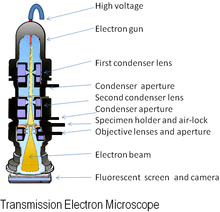
An electron microscope is a microscope that uses a beam of accelerated electrons as a source of illumination. As the wavelength of an electron can be up to 100, 000 times shorter than that of visible light photons, electron microscopes have a resolving power than light microscopes and can reveal the structure of smaller objects. Electron microscopes have electron optical lens systems that are analogous to the glass lenses of an optical light microscope.The similarity of scanning electron microscope (SEM) and transmission electron microscope (TEM) is are both structures smaller than 0.2 micrometer. The differences between of SEM and TEM are SEM produces a realistics 3D image of specimens's surface features but produces 2D image. The magnification of SEM is 1000X to 10,000X but in TEM are 10,000X to 100,000X. The resolving power for SEM is 20 nm but in TEM is 2.5 nm. SEM is to study the surface features of cells and viruses but TEM is to examine viruses or the internal ultrastructure in thin sections of cell. Electron Cryotomography is rapid technique provides way to preserve native state of structures examined in vacuum. Images recorded from many different directions to creates 3D structures.


Scanning Electron Microscope

view under electron microscope
         

Preparations of specimens for light microscopy 

Preparations of specimens for light microscopy are wet mount and smears. Smears-steps involve are preparing smears, fixation, and staining.Fixation is a preserves internal and external structures and fixed them in position. Stain is a salt composed of a positive and negative ion, one of which is colourful this will called the chromophore. Stain make internal and external structures of cell more visible by increasing contrast with background. There are three types of staining which are simple staining, differential staining and special staining. In differential staining have gram staining and acid-fast staining. In special staining have negative staining, endospore staining and flagella staining.

gram stain

acid-fast staining 

negative staining

endospore staining 

flagella staining 


Classification of organisms 

Taxonomy is a arrangement into groups based on mutual similarities. There are three interrelated parts in the taxonomy which classification, identification and nomenclature. Classification is the arrangement of organisms into groups or taxon based on mutual similarity or evolutionary relatedness. Identification is the process of discovering and recording the traits of organisms. Nomenclature is a assignment of names to taxonomic groups in agreement with published rules.Strain is a subgroup of species with one or more characteristics that distinguish it from other subgroups of the same species. Different ways to describe strains within a species which are biovars, morphovars and serovars. Phylogenetic classifications is a natural systems based on evolutionary relationships and direct comparison of genetic material and gene products.

Methods of classifying and identifying microorganisms

Methods of classifying and identifying microorganisms are morphological characteristics, differential staining, biochemical tests, serology, phage typing, fatty acid profiles, DNA base composition,DNA fingerprinting, nucleic acid hybridization. Methods use to classify and identify microorganisms after various analyses which are dichotomous keys and cladograms.


Microbiology Semester 2 Week 14

              Assalamualaikum and hi everyone... how are you all? I hope everyone will be fine as well... On Tuesday, before our class start...